Review
. 2015 Aug 19;370(1675):20140288.
doi: 10.1098/rstb.2014.0288.

The within-host dynamics of African trypanosome infections

Affiliations
Review

The within-host dynamics of African trypanosome infections

Keith R Matthews et al. Philos Trans R Soc Lond B Biol Sci. .

Abstract

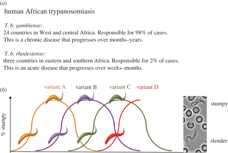
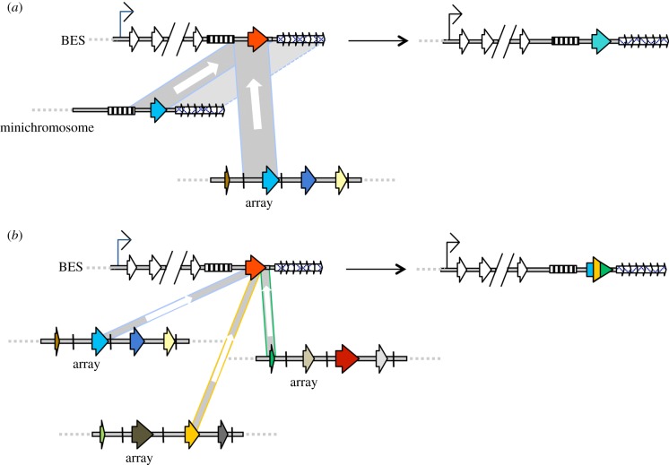
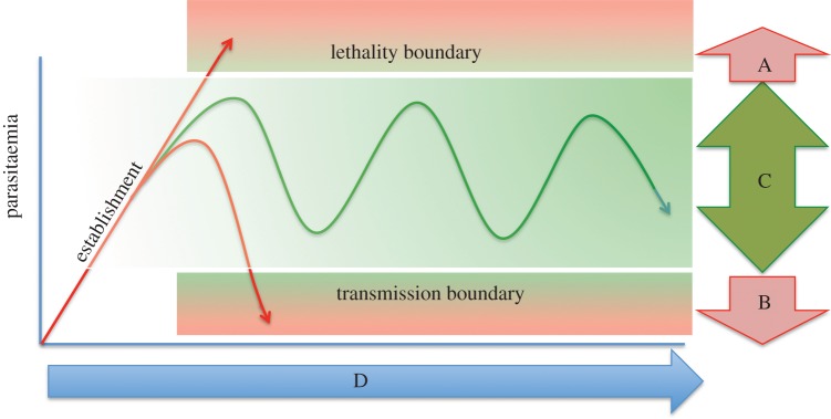
African trypanosomes are single-celled protozoan parasites that are capable of long-term survival while living extracellularly in the bloodstream and tissues of mammalian hosts. Prolonged infections are possible because trypanosomes undergo antigenic variation-the expression of a large repertoire of antigenically distinct surface coats, which allows the parasite population to evade antibody-mediated elimination. The mechanisms by which antigen genes become activated influence their order of expression, most likely by influencing the frequency of productive antigen switching, which in turn is likely to contribute to infection chronicity. Superimposed upon antigen switching as a contributor to trypanosome infection dynamics is the density-dependent production of cell-cycle arrested parasite transmission stages, which limit the infection while ensuring parasite spread to new hosts via the bite of blood-feeding tsetse flies. Neither antigen switching nor developmental progression to transmission stages is driven by the host. However, the host can contribute to the infection dynamic through the selection of distinct antigen types, the influence of genetic susceptibility or trypanotolerance and the potential influence of host-dependent effects on parasite virulence, development of transmission stages and pathogenicity. In a zoonotic infection cycle where trypanosomes circulate within a range of host animal populations, and in some cases humans, there is considerable scope for a complex interplay between parasite immune evasion, transmission potential and host factors to govern the profile and outcome of infection.

Keywords: Trypanosoma; antigenic variation; quorum sensing; transmission; trypanosome; within-host dynamics.


Figures

Figure 1.
Figure 1.
An overview of African trypanosomiaisis. (a) Shows a summary of the distribution and disease profile of the two species of African trypanosome responsible for human infection. Animal African trypanosomiasis (caused by T. brucei, T. congolense and T. vivax) is distributed throughout sub-Saharan Africa coincident with the distribution of the disease vector, tsetse flies. (b) Shows the conventional view of a trypanosome infection profile. Infection chronicity is achieved by appearance of a progression of waves of parasitaemia with distinct waves being composed of trypanosomes with antigenically distinct coats (for simplicity, each wave is shown as a single VSG, though normally many VSGs are represented per wave). Within each wave of parasitaemia, a developmental switch occurs, whereby proliferative slender forms become arrested stumpy forms as parasite numbers increase in response to the accumulation of the quorum-sensing signal, SIF.
Figure 2.
Figure 2.
VSG gene conversion during antigenic variation. (a) Gene conversion of intact VSG genes into the active bloodstream VSG expression site (BES), using donor VSGs that are either present in a minichromosome or a subtelomeric VSG array. In all cases, the VSGs are shown as coloured arrows and the extent of sequence copied during gene conversion is indicated by a grey box, within which the direction of copying is shown by a white arrow. During gene conversion from a silent minichromosome or array VSG (both light blue), the upstream boundary is normally 70 bp repeats (hatched box), which are found adjacent to most VSGs (though their numbers are lower when associated with array VSGs). The downstream boundary of gene conversion is frequently the 3′ end or 3′ flank of the VSG, though this can extend further when minichromosome VSGs act as donors, including reactions that encompass the telomere repeats (small, arrayed arrows). In the BES, ESAG genes (white arrows) are co-transcribed with the VSG (red) from a common promoter (thin black arrow); silent BES (not shown) can also act as donors of new VSGs by gene conversion, or can elicit VSG coat changes by transcriptional switching (not shown; see text). (b) Segmental gene conversion to form novel mosaic VSGs. In this reaction, multiple silent VSGs (here, three: light blue, orange and green) are recombined together to form a new VSG that is a composite of the three gene sequences. The VSGs that act as donors in segmental gene conversion are frequently pseudogenes and are normally located in disparate regions of the subtelomeric array archive. Many details of this reaction are uncertain, and some assumptions or simplifications are made: segmental gene conversion to form VSG mosaics may not happen in the active BES, as shown here; gene conversion is shown to encompass only VSG ORF-internal sequences, but the reactions may be ‘anchored’ by upstream or downstream homology; VSG mosaics normally display much greater intermingling of the donor VSG sequences than is indicated here.
Figure 3.
Figure 3.
The classical description of the interplay between antigenic variation and infection chronicity (redrawn from [58]). In scenario A, the parasites overgrow and kill the host. Scenario B occurs when parasites are rapidly cleared from the host. Scenario C is characteristic of trypanosome infections and is dependent upon both antigenic variation to evade specific immune responses (prevents scenario B) and density-dependent differentiation of slender to stumpy parasites (prevents scenario A). These processes are parasite-driven and independent of the host. Scenario C maximizes transmission, which will ultimately be the primary selective force on the trypanosome population. Several factors will determine the kinetics of infection in scenario C (i.e. infection duration and total parasite load; D) and these will include host susceptibility, parasite virulence and population factors such as herd immunity and co-infections, and the interplay of these factors with parasite antigenic variation and differentiation. These selective factors will shape the usage and evolution of the VSG repertoire at the individual and population levels.
Figure 4.
Figure 4.
Possible conflicts driving parasite virulence in different host settings. (a) The host range of different trypanosome species is shown, with T. b gambiense and T. b. rhodesiense being human infective. Trypanosoma brucei gambiense can also infect livestock, though human infection is more frequently detected; T. b. rhodesiense is most frequently found in livestock and game animals. Trypanosoma brucei brucei, T. congolense and T. vivax cannot infect humans but are maintained in game animals and livestock. (b) Three scenarios where parasites are transmitted either to trypanotolerant or susceptible hosts, or humans. In each scenario, potential outcomes are: (i) in trypanotolerant hosts, host suppression selects for increased virulence of the parasite population; (ii) parasites might exhibit increased virulence once released from either host suppression in susceptible animals or competition from co-infecting strains; and (iii) in humans, T. b. rhodesiense or T. b. gambiense are released from inter-species competition and may exhibit increased virulence.

References

    1. Brun R, Blum J, Chappuis F, Burri C. 2010. Human African trypanosomiasis. Lancet 375, 148–159. (10.1016/S0140-6736(09)60829-1) - DOI - PubMed
    1. Shaw AP, Cecchi G, Wint GRW, Mattioli RC, Robinson TP. 2014. Mapping the economic benefits to livestock keepers from intervening against bovine trypanosomosis in Eastern Africa. Prev. Vet. Med. 113, 197–210. (10.1016/j.prevetmed.2013.10.024) - DOI - PubMed
    1. Peacock L, Bailey M, Carrington M, Gibson W. 2014. Meiosis and haploid gametes in the pathogen Trypanosoma brucei. Curr. Biol. 24, 181–186. (10.1016/j.cub.2013.11.044) - DOI - PMC - PubMed
    1. Horn D. 2014. Antigenic variation in African trypanosomes. Mol. Biochem. Parasitol. 195, 123–129. (10.1016/j.molbiopara.2014.05.001) - DOI - PMC - PubMed
    1. Glover L, Hutchinson S, Alsford S, McCulloch R, Field MC, Horn D. 2013. Antigenic variation in African trypanosomes: the importance of chromosomal and nuclear context in VSG expression control. Cell Microbiol. 15, 1984–1993. (10.1111/cmi.12215) - DOI - PMC - PubMed

Publication types

MeSH terms

Substances

LinkOut - more resources