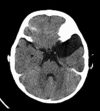
Spontaneous Rupture of the Middle Fossa Arachnoid Cyst into the Subdural Space: Case Report
- PMID: 26150904
- PMCID: PMC4482331
- DOI: 10.12659/PJR.893928
Spontaneous Rupture of the Middle Fossa Arachnoid Cyst into the Subdural Space: Case Report
Abstract
Background: Arachnoid cysts are congenital, benign and intra-arachnoidal lesions. A great majority of arachnoid cysts are congenital. However, to a lesser extent, they are known to develop after head trauma and brain inflammatory diseases. Arachnoid cysts are mostly asymptomatic and they can develop anywhere in the brain along the arachnoid membrane.
Case report: Arachnoid cysts form 1% of the non-traumatic lesions which occupy a place and it is thought to be a congenital lesion developed as a result of meningeal development abnormalities or a lesion acquired after trauma and infection. There is a male dominance at a rate of 3/1 in arachnoid cysts which locate mostly in the middle fossa. Our patient was a 2-years-old boy.
Conclusions: As a conclusion, spontaneous subdural hygroma is a rare complication of the arachnoid cysts. Surgical intervention could be required in acute cases.
Keywords: Arachnoid Cysts; Magnetic Resonance Imaging; Subdural Effusion; Tomography Scanners, X-Ray Computed.
Figures


Similar articles
-
Spontaneous arachnoid cyst rupture in a previously asymptomatic child: a case report.Eur J Paediatr Neurol. 2004;8(5):247-51. doi: 10.1016/j.ejpn.2004.04.005. Eur J Paediatr Neurol. 2004. PMID: 15341907
-
Spontaneous Subdural Haematoma Developing Secondary to Arachnoid Cyst Rupture.J Clin Diagn Res. 2016 Oct;10(10):PD05-PD06. doi: 10.7860/JCDR/2016/21056.8708. Epub 2016 Oct 1. J Clin Diagn Res. 2016. PMID: 27891397 Free PMC article.
-
Arachnoid cyst rupture producing subdural hygroma and intracranial hypertension: case reports.Neurosurgery. 1997 Oct;41(4):951-5; discussion 955-6. doi: 10.1097/00006123-199710000-00036. Neurosurgery. 1997. PMID: 9316060 Review.
-
Subdural hygroma after spontaneous rupture of an arachnoid cyst in a pediatric patient: A case report.Radiol Case Rep. 2020 Dec 1;16(2):309-311. doi: 10.1016/j.radcr.2020.11.036. eCollection 2021 Feb. Radiol Case Rep. 2020. PMID: 33304441 Free PMC article.
-
Arachnoid cyst rupture with subdural hygroma: report of three cases and literature review.Childs Nerv Syst. 2002 Nov;18(11):609-13. doi: 10.1007/s00381-002-0651-7. Epub 2002 Aug 29. Childs Nerv Syst. 2002. PMID: 12420120 Review.
Cited by
-
Subdural Lesions Linking Additional Intracranial Spaces and Chronic Subdural Hematomas: A Narrative Review with Mutual Correlation and Possible Mechanisms behind High Recurrence.Diagnostics (Basel). 2023 Jan 8;13(2):235. doi: 10.3390/diagnostics13020235. Diagnostics (Basel). 2023. PMID: 36673045 Free PMC article. Review.
-
Spontaneous chronic subdural hematoma associated with arachnoid cyst in a child: A case report and critical review of the literature.Surg Neurol Int. 2022 Apr 15;13:156. doi: 10.25259/SNI_100_2022. eCollection 2022. Surg Neurol Int. 2022. PMID: 35509549 Free PMC article.
-
Ruptured Sylvian arachnoid cysts: an update on a real problem.Childs Nerv Syst. 2023 Jan;39(1):93-119. doi: 10.1007/s00381-022-05685-3. Epub 2022 Sep 28. Childs Nerv Syst. 2023. PMID: 36169701 Free PMC article. Review.
-
Multiple etiologies of secondary headaches associated with arachnoid cyst, cerebrospinal fluid hypovolemia, and nontraumatic chronic subdural hematoma in an adolescent: A case report.Surg Neurol Int. 2022 Aug 26;13:386. doi: 10.25259/SNI_327_2022. eCollection 2022. Surg Neurol Int. 2022. PMID: 36128159 Free PMC article.
-
Spontaneous rupture of an arachnoid cyst in an adult: illustrative case.J Neurosurg Case Lessons. 2023 Feb 13;5(7):CASE22420. doi: 10.3171/CASE22420. Print 2023 Feb 13. J Neurosurg Case Lessons. 2023. PMID: 38015025 Free PMC article.
References
-
- Görücü Y, Barut AY, Mutlu İN: Araknoid kist (olgu sunumu) İstanbul Tıp Dergisi. 2006;4:43–46. [in Turkish]
-
- Hatipoğlu MA, et al. Arachnoid cyst ruptures into subdural space after head trauma: case report. Türk Nöroşirürji Dergisi. 2005;15(1):76–78. [in Turkish]
-
- Garcia Santos JM, Martinez-Lage J, Gilabert Ubeda A, et al. Arachnoid cysts of the middle cranial fossa: a consideration of their origins based on imaging. Neuroradiology. 1993;35:355–58. - PubMed
-
- Poirrier Anne-Lise ML, Misson J, et al. Spontaneous arachnoid cyst rupture in a previously asymptomatic child: a case report. Eur J Paediatr Neurol. 2004;8:247–51. - PubMed
-
- Gosalakkal J. Intracranial arachnoid cysts in children: a review of pathogenesis, clinical features, and management. Pediatr Neurol. 2002;26:93–98. - PubMed
Publication types
LinkOut - more resources
Full Text Sources
Research Materials
