. 2015 Jul 7:6:7623.
doi: 10.1038/ncomms8623.

Recessive mutations in POLR1C cause a leukodystrophy by impairing biogenesis of RNA polymerase III

Affiliations

Recessive mutations in POLR1C cause a leukodystrophy by impairing biogenesis of RNA polymerase III

Isabelle Thiffault et al. Nat Commun. .

Abstract

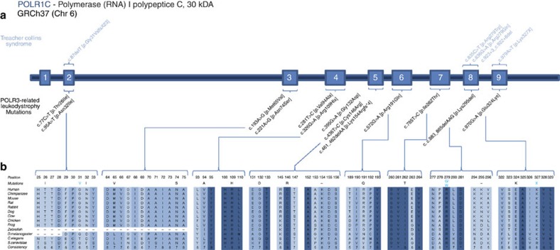
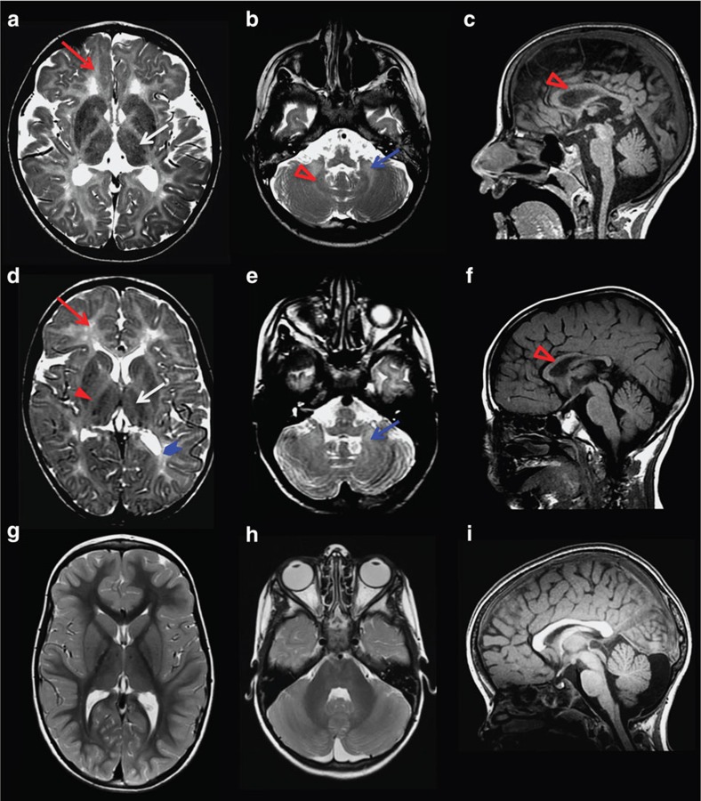
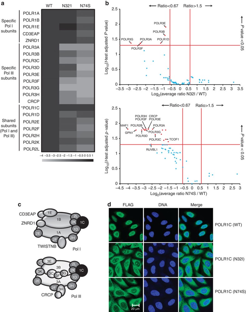
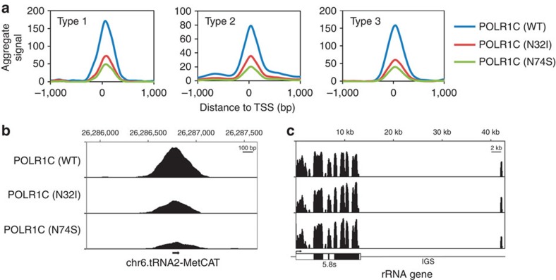
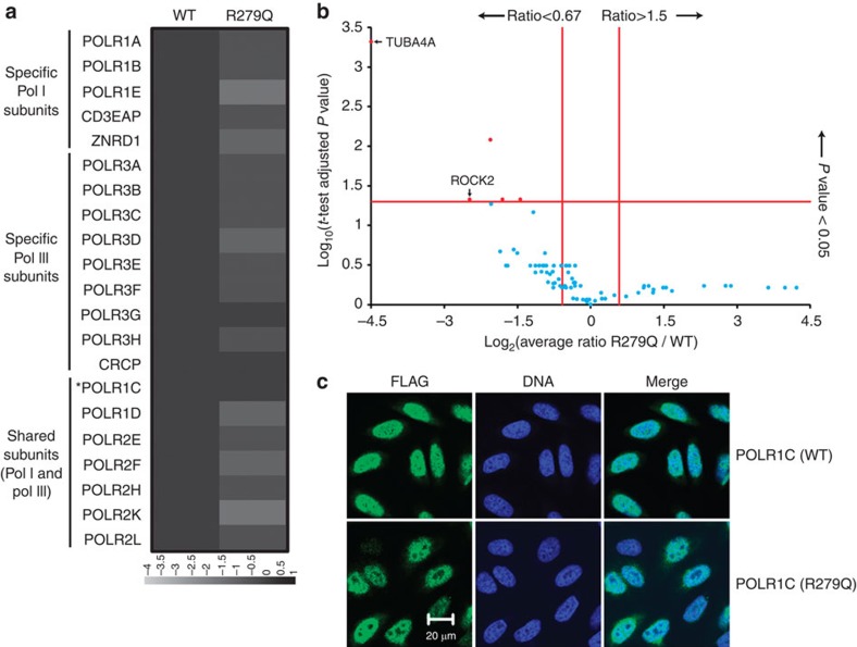
A small proportion of 4H (Hypomyelination, Hypodontia and Hypogonadotropic Hypogonadism) or RNA polymerase III (POLR3)-related leukodystrophy cases are negative for mutations in the previously identified causative genes POLR3A and POLR3B. Here we report eight of these cases carrying recessive mutations in POLR1C, a gene encoding a shared POLR1 and POLR3 subunit, also mutated in some Treacher Collins syndrome (TCS) cases. Using shotgun proteomics and ChIP sequencing, we demonstrate that leukodystrophy-causative mutations, but not TCS mutations, in POLR1C impair assembly and nuclear import of POLR3, but not POLR1, leading to decreased binding to POLR3 target genes. This study is the first to show that distinct mutations in a gene coding for a shared subunit of two RNA polymerases lead to selective modification of the enzymes' availability leading to two different clinical conditions and to shed some light on the pathophysiological mechanism of one of the most common hypomyelinating leukodystrophies, POLR3-related leukodystrophy.


Figures

Figure 1
Figure 1. POLR1C mutations in leukodystrophy and TCS cases.
(a) Genomic organization of POLR1C in humans (UCSC Genome Browser hg19): mutations and their positions within the POLR1C gDNA; in light blue are mutations that cause TCS, mutations in black cause POLR3-related leukodystrophy. (b) POLR1C mutations in patients with leukodystrophy affect amino acids that are conserved through species.
Figure 2
Figure 2. MRI characteristics of POLR3-related leukodystrophy caused by POLR1C mutations.
Axial T2-weighted (a,b,d,e,g,h) and sagittal T1-weighted (c,f,i) images of case 1 aged 6 years (a–c) and case 2 aged 4.5 years (d–f) compared with a healthy control aged 4 years (g–i). Diffuse hyperintense signal of the supratentorial (red arrow, a,d) and cerebellar (blue arrow, b,e) white matter is visible on the T2-weighted images, indicating hypomyelination. There is no cerebellar atrophy. As typical for POLR3-related leukodystrophy, the ventrolateral thalamus (white arrow, a,d), the optic radiation (thick arrowhead blue, d) and the dentate nucleus (open red arrowhead, b) show a relative hypointense signal on the T2-weighted images resulting in an easily visible dentate nucleus (b) as compared with the control (h) as well as a small dot in the posterior limb of the internal capsule (red arrowhead, d). The corpus callosum is slightly thinned in case 1 and thinned in case 2 (open red arrowhead, c,f).
Figure 3
Figure 3. Impact of POLR1C mutations on polymerase assembly and nuclear import.
(a) FLAG-tagged POLR1C variants, either the wild-type (1C) polypeptide or mutated versions having a N32I or a N74S substitution, were expressed in HeLa cells and purified using anti-FLAG affinity chromatography. The co-purified proteins were identified using LC-MS/MS mass spectrometry. The heatmap contains the log2-transformed average spectral count ratios N32I or N74S/WT across all three replicates. Spectral counts were computed with Mascot (see Supplementary Table 6 for the complete data set). Specific and shared POLR1 (Pol I) and POLR3 (Pol III) subunits are identified on the left. POLR1C (the bait) is identified by an asterisk. (b) Volcano plots of the log2-transformed average spectral count ratios N32I or N74S/WT (x axis) and the –log10-transformed P values (adjusted with the Benjamini–Hochberg procedure) resulting from the two-tailed one-sample t-tests of the high-confidence interactors of POLR1C. Red proteins show a level of differential interaction with POLR1C that is statistically significant, while blue proteins do not. (c) Schematic representation of the subunit composition of POLR1 (Pol I) and POLR3 (Pol III; see refs for details). Shared subunits are in grey and POLR1C in black. (d) Immunofluorescence experiments showing the cellular localization of tagged POLR1C variants. Nuclei are stained using TO-PRO-3 iodine. Scale bar, 20 μm.
Figure 4
Figure 4. Impact of POLR1C mutations on polymerase association with chromatin.
(a) ChIP-Seq experiments of FLAG-tagged POLR1C variants (wild type, N32I or N74S). Aggregate profile produced with the annotation mode of the Versatile Aggregate Profiler shows ChIP-Seq data sets over the three classes of POLR3-transcribed genes, as defined by the promoter structure. Type 1 genes have an internal promoter composed of A and C boxes (5S rRNA genes). Type 2 genes have an internal promoter composed of A and B boxes (tRNA genes, for example), while the promoter of type 3 genes (U6, 7SK, RNase P and others) is located upstream of the transcription start site (TSS). The TSS was used as the reference point. (b) IGV view of a tRNA-Met gene transcribed by POLR3. POLR1C binding is decreased in mutated variants compared with wild type. IGS, intergenic spacer. (c) IGV view of one ribosomal DNA (rDNA) repeat. The rDNA gene encodes a 45S pre-rRNA precursor that will generate the 5.8S, 18S and 28S rRNAs. There are ∼400 copies of the rDNA gene arranged in tandem repeats in the human genome. rDNA repeats are not present in the reference genome assemblies; therefore, unique reads were aligned directly to the human rDNA reference sequence (NCBI accession number: HSU13369). No differences were observed in POLR1C occupancy between wild-type and mutant variants. A schematic of a rDNA repeat is included below the graph.
Figure 5
Figure 5. Impact of a TCS-causative mutation in POLR1C on polymerase assembly and cellular localization.
FLAG-tagged POLR1C variants, either the wild type (1C) or a mutated version with the R279Q substitution, were expressed, affinity-purified and used in anti-FLAG immunofluorescence experiments as in Fig. 3. (a) Affinity purification coupled to mass spectrometry data is represented in the form of a heatmap that contains the log2-transformed average spectral count ratios R279Q/WT across all three replicates. Spectral counts were computed with Mascot (see legend to Fig. 3 for details). (b) Volcano plot of the log2-transformed average spectral count ratios N279Q/WT (x axis) and the –log10-transformed P-values (adjusted with the Benjamini–Hochberg procedure) resulting from the two-tailed one-sample t-tests of the high-confidence interactors of POLR1C. Red proteins show a level of differential interaction with POLR1C that is statistically significant, while blue proteins do not. Proteins with a log2-transformed average spectral count ratio <−4.5 were capped to −4.5 for display purposes. (c) Immunofluorescence data showing the cellular localization of tagged POLR1C variants are shown. Scale bar, 20 μm.

Similar articles

  • The leukodystrophy mutation Polr3b R103H causes homozygote mouse embryonic lethality and impairs RNA polymerase III biogenesis.
    Choquet K, Pinard M, Yang S, Moir RD, Poitras C, Dicaire MJ, Sgarioto N, Larivière R, Kleinman CL, Willis IM, Gauthier MS, Coulombe B, Brais B. Choquet K, et al. Mol Brain. 2019 Jun 20;12(1):59. doi: 10.1186/s13041-019-0479-7. Mol Brain. 2019. PMID: 31221184 Free PMC article.
  • Craniofacial features of POLR3-related leukodystrophy caused by biallelic variants in POLR3A, POLR3B and POLR1C.
    Mirchi A, Guay SP, Tran LT, Wolf NI, Vanderver A, Brais B, Sylvain M, Pohl D, Rossignol E, Saito M, Moutton S, González-Gutiérrez-Solana L, Thiffault I, Kruer MC, Moron DG, Kauffman M, Goizet C, Sztriha L, Glamuzina E, Melançon SB, Naidu S, Retrouvey JM, Lacombe S, Bernardino-Cuesta B, De Bie I, Bernard G. Mirchi A, et al. J Med Genet. 2023 Oct;60(10):1026-1034. doi: 10.1136/jmg-2023-109223. Epub 2023 May 16. J Med Genet. 2023. PMID: 37197783 Free PMC article.
  • Endocrine and Growth Abnormalities in 4H Leukodystrophy Caused by Variants in POLR3A, POLR3B, and POLR1C.
    Pelletier F, Perrier S, Cayami FK, Mirchi A, Saikali S, Tran LT, Ulrick N, Guerrero K, Rampakakis E, van Spaendonk RML, Naidu S, Pohl D, Gibson WT, Demos M, Goizet C, Tejera-Martin I, Potic A, Fogel BL, Brais B, Sylvain M, Sébire G, Lourenço CM, Bonkowsky JL, Catsman-Berrevoets C, Pinto PS, Tirupathi S, Strømme P, de Grauw T, Gieruszczak-Bialek D, Krägeloh-Mann I, Mierzewska H, Philippi H, Rankin J, Atik T, Banwell B, Benko WS, Blaschek A, Bley A, Boltshauser E, Bratkovic D, Brozova K, Cimas I, Clough C, Corenblum B, Dinopoulos A, Dolan G, Faletra F, Fernandez R, Fletcher J, Garcia Garcia ME, Gasparini P, Gburek-Augustat J, Gonzalez Moron D, Hamati A, Harting I, Hertzberg C, Hill A, Hobson GM, Innes AM, Kauffman M, Kirwin SM, Kluger G, Kolditz P, Kotzaeridou U, La Piana R, Liston E, McClintock W, McEntagart M, McKenzie F, Melançon S, Misbahuddin A, Suri M, Monton FI, Moutton S, Murphy RPJ, Nickel M, Onay H, Orcesi S, Özkınay F, Patzer S, Pedro H, Pekic S, Pineda Marfa M, Pizzino A, Plecko B, Poll-The BT, Popovic V, Rating D, Rioux MF, Rodriguez Espinosa N, Ronan A, Ostergaard JR, Rossignol E, Sanchez-Carpintero R, Schossig A, Senbil N, Sønderberg Roos LK, Stevens CA, Synofzik M, S… See abstract for full author list ➔ Pelletier F, et al. J Clin Endocrinol Metab. 2021 Jan 23;106(2):e660-e674. doi: 10.1210/clinem/dgaa700. J Clin Endocrinol Metab. 2021. PMID: 33005949 Free PMC article.
  • POLR3-related leukodystrophy: How do mutations affecting RNA polymerase III subunits cause hypomyelination?
    Coulombe B, Derksen A, La Piana R, Brais B, Gauthier MS, Bernard G. Coulombe B, et al. Fac Rev. 2021 Feb 5;10:12. doi: 10.12703/r/10-12. eCollection 2021. Fac Rev. 2021. PMID: 33659930 Free PMC article. Review.
  • Autosomal recessive Treacher Collins syndrome due to POLR1C mutations: Report of a new family and review of the literature.
    Ghesh L, Vincent M, Delemazure AS, Boyer J, Corre P, Perez F, Geneviève D, Laplanche JL, Collet C, Isidor B. Ghesh L, et al. Am J Med Genet A. 2019 Jul;179(7):1390-1394. doi: 10.1002/ajmg.a.61147. Epub 2019 Apr 8. Am J Med Genet A. 2019. PMID: 30957429 Review.

Cited by

References

    1. Schiffmann R. & van der Knaap M. S. The latest on leukodystrophies. Curr. Opin. Neurol. 17, 187–192 (2004). - PubMed
    1. Schiffmann R. & van der Knaap M. S. Invited article: an MRI-based approach to the diagnosis of white matter disorders. Neurology 72, 750–759 (2009). - PMC - PubMed
    1. Bernard G. & Vanderver A. in GeneReviews® (eds Pagon, R. A., Adam, M. P., Ardinger, H. H., et al..) (University of Washington, Seattle, 1993–2015), http://www.ncbi.nlm.nih.gov/books/NBK99167/. - PubMed
    1. Bernard G. et al.. Mutations of POLR3A encoding a catalytic subunit of RNA polymerase Pol III cause a recessive hypomyelinating leukodystrophy. Am. J. Hum. Genet. 89, 415–423 (2011). - PMC - PubMed
    1. Tetreault M. et al.. Recessive mutations in POLR3B, encoding the second largest subunit of Pol III, cause a rare hypomyelinating leukodystrophy. Am. J. Hum. Genet. 89, 652–655 (2011). - PMC - PubMed

Publication types

MeSH terms

Associated data