. 2015 Jul 7:6:7499.
doi: 10.1038/ncomms8499.

SUMO and ubiquitin-dependent XPC exchange drives nucleotide excision repair

Affiliations

SUMO and ubiquitin-dependent XPC exchange drives nucleotide excision repair

Loes van Cuijk et al. Nat Commun. .

Abstract

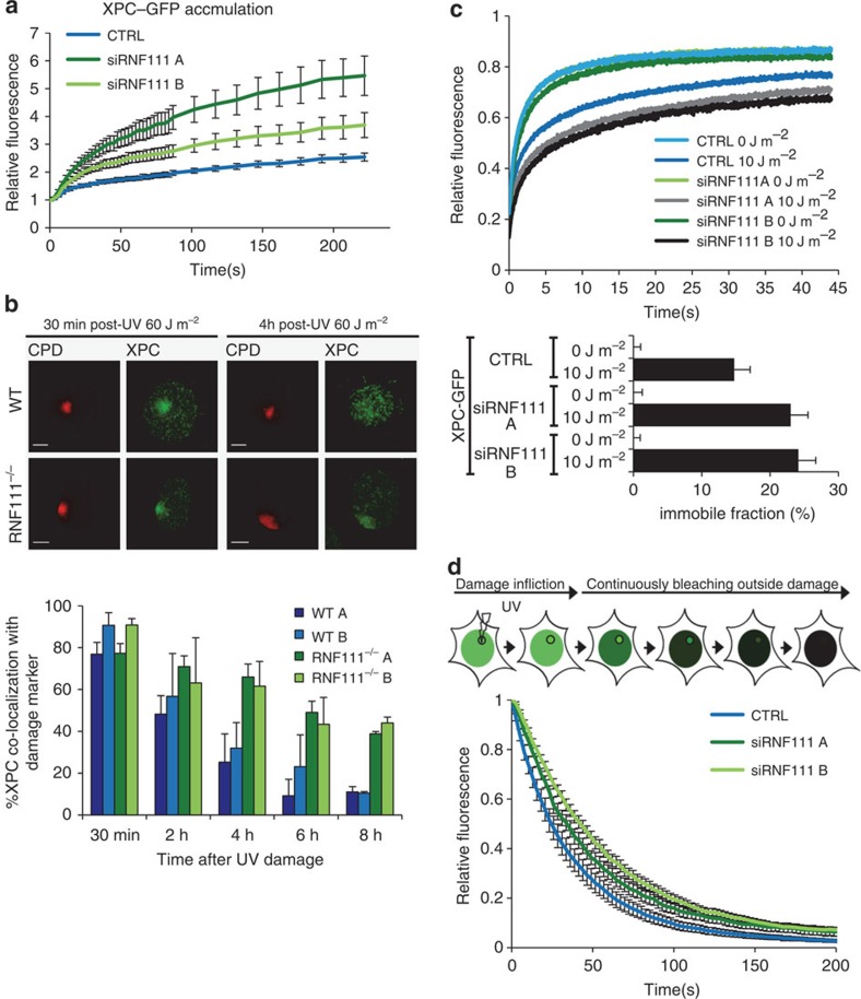
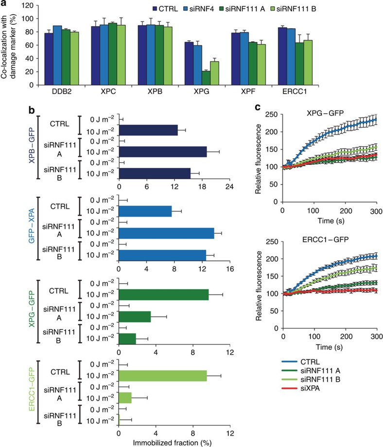
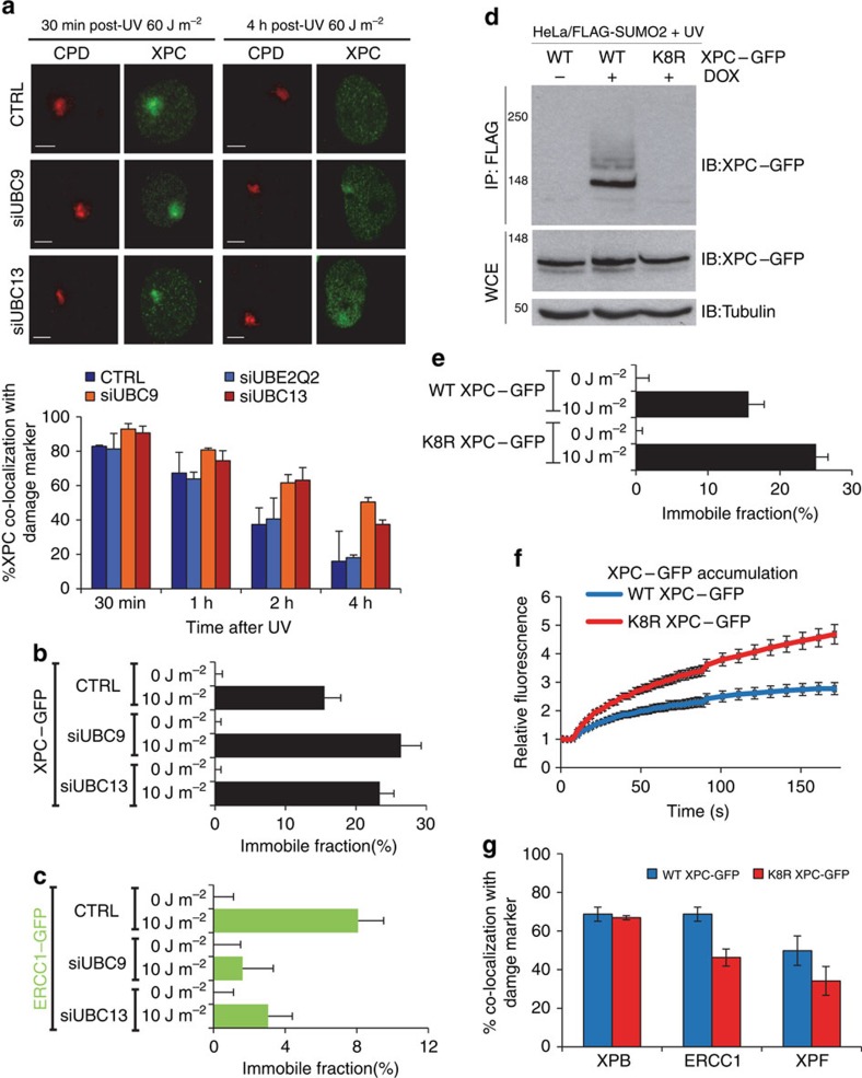
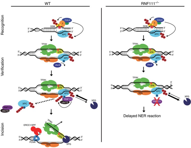
XPC recognizes UV-induced DNA lesions and initiates their removal by nucleotide excision repair (NER). Damage recognition in NER is tightly controlled by ubiquitin and SUMO modifications. Recent studies have shown that the SUMO-targeted ubiquitin ligase RNF111 promotes K63-linked ubiquitylation of SUMOylated XPC after DNA damage. However, the exact regulatory function of these modifications in vivo remains elusive. Here we show that RNF111 is required for efficient repair of ultraviolet-induced DNA lesions. RNF111-mediated ubiquitylation promotes the release of XPC from damaged DNA after NER initiation, and is needed for stable incorporation of the NER endonucleases XPG and ERCC1/XPF. Our data suggest that RNF111, together with the CRL4(DDB2) ubiquitin ligase complex, is responsible for sequential XPC ubiquitylation, which regulates the recruitment and release of XPC and is crucial for efficient progression of the NER reaction, thereby providing an extra layer of quality control of NER.


Figures

Figure 1
Figure 1. RNF111 is necessary for efficient GG-NER.
(a) Representative pictures of unscheduled DNA synthesis (UDS) of the indicated MEFs, determined by 5-ethynyl-2′-deoxyuridine (EdU) incorporation over 3 h after UV irradiation (16 J m−2). Scale bar, 25 μm. (b) Quantification of UDS levels in MEFs, as determined by EdU incorporation over a time period of 3 or 9 h after UV irradiation (16 J m−2). UDS levels in WT MEFs were set at 100% (n>100 cells per sample, in at least two independent experiment; error bars are the mean±s.d.). (c) 6-4PP removal assayed by immunofluorescence, using a 6-4PP specific antibody. The indicated MEFs were UV-irradiated (10 J m−2) and allowed to repair 6-4PPs for the indicated time points. Relative fluorescence directly after UV exposure was set at 100%. (n>70 cells, three independent experiments; error bars are the mean±s.d.).
Figure 2
Figure 2. RNF111 is required for XPC release.
(a) Relative XPC–GFP accumulation at sites of LUD in control and RNF111-depleted cells. GFP fluorescence intensity at UV-C laser induced LUD was measured over time using live-cell confocal imaging and quantified to predamage intensity set at 1 at t=0 (n>15 cells per sample, measured in two independent experiments; error bars are the mean±2 × s.e.m.). (b) Top panel: representative immunofluorescence pictures of co-localization of XPC with CPD at LUD in WT and Rnf111−/−MEFs at the indicated time points after UV irradiation (60 J m−2) are shown. Scale bars, 5 μm. Lower panel: quantification of the XPC co-localization with CPD (n>50 cells with LUD were analysed per sample in three independent experiments; error bars are the mean±s.d.). (c) Top panel: FRAP analysis of XPC–GFP in mock treated or global UV-irradiated (10 J m−2) XP4PA (XPC deficient) cells, on transfection with the indicated siRNA's. XPC–GFP was bleached in a small strip within the nucleus and fluorescence recovery was measured over 45 s and normalized to prebleach intensity (n=40; from two independent experiments error bars are the mean±2 × s.e.m.). The immobilized fraction (%)=1−((average fluorescence intensity UV-irradiated cells−the first data point after bleaching)/(average fluorescence intensity unchallenged cells−the first data point after bleaching)), is plotted in the lower panel. The immobilized fraction was calculated over the last 10 s. (d) Inverse FRAP (iFRAP) analysis of XPC–GFP at LUD. XP4PA cells stably expressing XPC–GFP were transfected with the indicated siRNA's. Seventy-two hours after transfection, cells were locally exposed to a 266-nm UV-C laser. After the accumulation plateau was reached (5 min after exposure) the undamaged part of the nucleus was continuously bleached and fluorescence in the damaged area was monitored. Fluorescence was normalized to prebleach intensity (n>15 cells per sample, measured in two independent experiments; error bars are the mean±s.e.m.).
Figure 3
Figure 3. RNF111 is required for binding of XPG and XPF/ERCC1 to the NER complex.
(a) U2OS cells expressing CPD–photolyase–mCherry were transfected with the indicated siRNA's 3 days before the immunofluorescence experiment. Cells were local UV irradiated (60 J m−2) and immunostained for the indicated proteins 30 min later. The percentage of co-localization with the damage marker CPD–photolyase–mCherry at LUD is plotted in the graph (n>50 cells containing a LUD were scored in at least three independent experiments; error bars are the mean±s.d.). (b) The immobilized fraction of XPB–GFP, GFP–XPA, XPG–GFP and ERCC1–GFP as determined by FRAP analysis in mock or UV-treated (10 J m−2) cells on transfection with the indicated siRNA's (n>32 cells from at least two independent experiments; error bars are the mean±2 × s.e.m.). (c) Cells stably expressing XPG–GFP and ERCC1–GFP transfected with the indicated siRNA's were locally irradiated using a 266-nm UV-C laser. GFP fluorescence intensity at LUD was monitored for 6 min, with 10 s intervals and normalized to predamage values. (n=24 cells from three independent experiments; error bars are the mean±s.e.m.).
Figure 4
Figure 4. XPC release is SUMO and K63-ubiquitylation dependent.
(a) Top panel: representative pictures of co-localization of XPC with CPD at LUD in U2OS cells transfected with the indicated siRNA's 30 min or 4 h after local UV irradiation (60 J m−2) are shown. Scale bar, 5 μm. Bottom panel: quantification of XPC co-localization with the damage marker CPD. (n≈50 cells containing a LUD were scored per sample in three independent experiments; error bars are the mean±s.d.). The immobilized fraction of XPC–GFP (b) or ERCC1–GFP (c) as determined by FRAP analysis in mock or UV-treated (10 J m−2) cells depleted by siRNA of UBC9 or UBC13 (n=40 from two experiments; error bars are the mean±2 × s.e.m.). (d) HeLa/FLAG-SUMO2 cells were transfected with plasmids expressing WT or K8R XPC–GFP, then left untreated or incubated with doxycycline (DOX) to induce FLAG-SUMO2 expression. One hour after UV exposure (16 J m−2), cells were lysed under denaturing conditions, and XPC SUMOylation was analysed by immunoblotting of FLAG IPs with GFP antibody. (e) The immobilized fraction of WT XPC–GFP or K8R XPC–GFP as determined by FRAP analysis in mock or UV-treated (10 J m−2) cells (n>40 from three experiments; error bars are the mean±2 × s.e.m.). (f) Cells stably expressing WT XPC–GFP or K8R XPC–GFP were locally irradiated using a 266 nm UV-C laser. GFP fluorescence intensity at UV-C laser-induced LUD was measured over time using live-cell confocal imaging and quantified to predamage intensity set at 1 at t=0 (n>25 cells per sample, measured in two independent experiments; error bars are the mean±s.e.m.). (g) XPC deficient XP4PA cells stably expressing WT XPC–GFP of K8R XPC–GFP were locally UV-irradiated (60 J m−2) and immunostained for endogenous XPB, ERCC1 and XPF proteins 30 min later. The percentage of co-localization with GFP–XPC at LUD is plotted in the graph (n>100 cells containing a LUD were scored in at two independent experiments; error bars are the mean±s.d.).
Figure 5
Figure 5. Proposed model for RNF111-dependent XPC ubiquitylation in controlling NER.
In WT cells SUMOylated XPC is ubiquitylated by RNF111, promoting its release from damaged DNA. This RNF111-mediated XPC release facilitates XPG and ERCC1/XPF recruitment, thereby enabling an efficient NER reaction. In RNF111−/− cells, where XPC is not modified by RNF111-dependent K63 ubiquitin chains, XPC remains more stably associated with the preincision NER complex. This interferes with proper loading of XPG, thereby inhibiting the NER reaction. Branched ‘Ub' represents K48-linked Ub chains, linear ‘Ub' represents K63-linked Ub chains; ‘S' represents SUMOylation; ‘1' and ‘2' represent DDB1 and DDB2, respectively.

References

    1. Marteijn J. A., Lans H., Vermeulen W. & Hoeijmakers J. H. Understanding nucleotide excision repair and its roles in cancer and ageing. Nat. Rev. Mol. Cell Biol. 15, 465–481 (2014). - PubMed
    1. Yokoi M. et al.. The xeroderma pigmentosum group C protein complex XPC-HR23B plays an important role in the recruitment of transcription factor IIH to damaged DNA. J. Biol. Chem. 275, 9870–9875 (2000). - PubMed
    1. Volker M. et al.. Sequential assembly of the nucleotide excision repair factors in vivo. Mol. Cell 8, 213–224 (2001). - PubMed
    1. Camenisch U., Dip R., Schumacher S. B., Schuler B. & Naegeli H. Recognition of helical kinks by xeroderma pigmentosum group A protein triggers DNA excision repair. Nat. Struct. Mol. Biol. 13, 278–284 (2006). - PubMed
    1. Sugasawa K., Akagi J., Nishi R., Iwai S. & Hanaoka F. Two-step recognition of DNA damage for mammalian nucleotide excision repair: directional binding of the XPC complex and DNA strand scanning. Mol. Cell 36, 642–653 (2009). - PubMed

Publication types

MeSH terms