

Genetic determinants of antithyroid drug-induced agranulocytosis by human leukocyte antigen genotyping and genome-wide association study
- PMID: 26151496
- PMCID: PMC4506516
- DOI: 10.1038/ncomms8633
Genetic determinants of antithyroid drug-induced agranulocytosis by human leukocyte antigen genotyping and genome-wide association study
Abstract
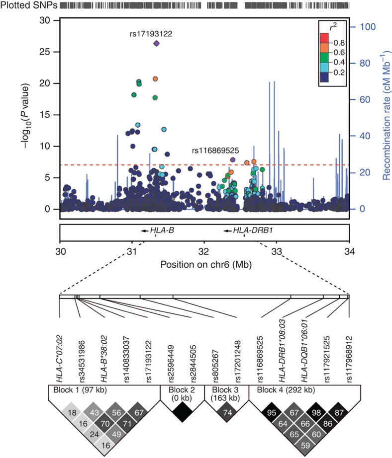
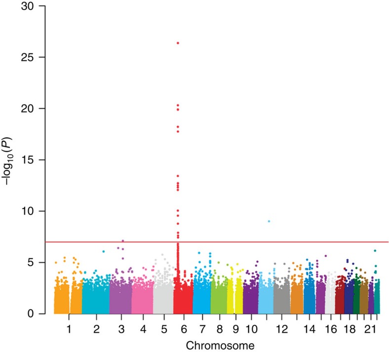
Graves' disease is the leading cause of hyperthyroidism affecting 1.0-1.6% of the population. Antithyroid drugs are the treatment cornerstone, but may cause life-threatening agranulocytosis. Here we conduct a two-stage association study on two separate subject sets (in total 42 agranulocytosis cases and 1,208 Graves' disease controls), using direct human leukocyte antigen genotyping and SNP-based genome-wide association study. We demonstrate HLA-B*38:02 (Armitage trend Pcombined=6.75 × 10(-32)) and HLA-DRB1*08:03 (Pcombined=1.83 × 10(-9)) as independent susceptibility loci. The genome-wide association study identifies the same signals. Estimated odds ratios for these two loci comparing effective allele carriers to non-carriers are 21.48 (95% confidence interval=11.13-41.48) and 6.13 (95% confidence interval=3.28-11.46), respectively. Carrying both HLA-B*38:02 and HLA-DRB1*08:03 increases odds ratio to 48.41 (Pcombined=3.32 × 10(-21), 95% confidence interval=21.66-108.22). Our results could be useful for antithyroid-induced agranulocytosis and potentially for agranulocytosis caused by other chemicals.
Figures
References
-
- Weetman A. P. Graves' disease. N. Engl. J. Med. 343, 1236–1248 (2000). - PubMed
-
- Tunbridge W. M. et al.. The spectrum of thyroid disease in a community: the Whickham survey. Clin. Endocrinol. (Oxf.) 7, 481–493 (1977). - PubMed
-
- Jacobson D. L., Gange S. J., Rose N. R. & Graham N. M. Epidemiology and estimated population burden of selected autoimmune diseases in the United States. Clin. Immunol. Immunopathol. 84, 223–243 (1997). - PubMed
-
- Cooper D. S. Antithyroid drugs. N. Engl. J. Med. 352, 905–917 (2005). - PubMed
-
- Tajiri J. & Noguchi S. Antithyroid drug-induced agranulocytosis: special reference to normal white blood cell count agranulocytosis. Thyroid 14, 459–462 (2004). - PubMed
Publication types
MeSH terms
Substances
LinkOut - more resources
Full Text Sources
Other Literature Sources
Molecular Biology Databases
Research Materials
