. 2015 Jul 6;34(1):73-84.
doi: 10.1016/j.devcel.2015.05.012.

Establishment of Centromeric Chromatin by the CENP-A Assembly Factor CAL1 Requires FACT-Mediated Transcription

Affiliations

Establishment of Centromeric Chromatin by the CENP-A Assembly Factor CAL1 Requires FACT-Mediated Transcription

Chin-Chi Chen et al. Dev Cell. .

Abstract

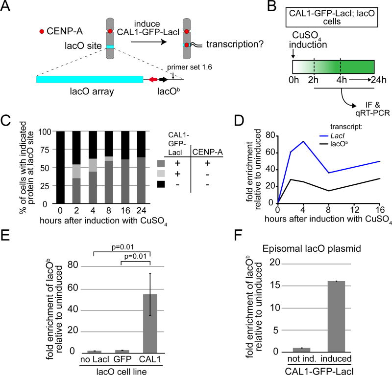
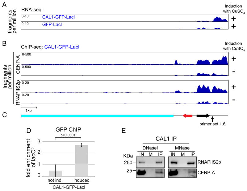
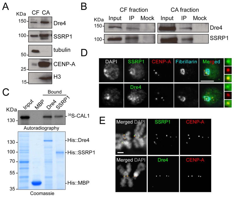
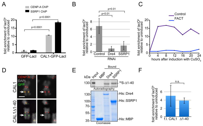
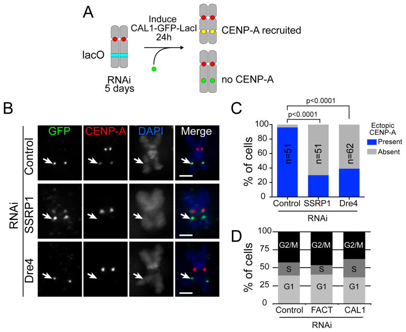
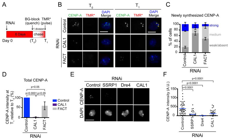
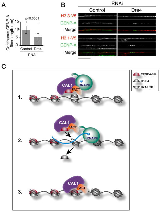
Centromeres are essential chromosomal structures that mediate accurate chromosome segregation during cell division. Centromeres are specified epigenetically by the heritable incorporation of the centromeric histone H3 variant CENP-A. While many of the primary factors that mediate centromeric deposition of CENP-A are known, the chromatin and DNA requirements of this process have remained elusive. Here, we uncover a role for transcription in Drosophila CENP-A deposition. Using an inducible ectopic centromere system that uncouples CENP-A deposition from endogenous centromere function and cell-cycle progression, we demonstrate that CENP-A assembly by its loading factor, CAL1, requires RNAPII-mediated transcription of the underlying DNA. This transcription depends on the CAL1 binding partner FACT, but not on CENP-A incorporation. Our work establishes RNAPII passage as a key step in chaperone-mediated CENP-A chromatin establishment and propagation.


Figures

Figure 1
Figure 1. CENP-A deposition at the ectopic lacO site is associated with transcription
A) Experimental approach to determine if transcription is coupled with CENP-A deposition. The lacO vector is stably inserted in S2 cells and contains 256 lacO repeats (lacO array; blue bar), the bacterial Amp resistance gene (black arrow), and the yeast TRP1 gene (red arrow). Primer set 1.6 (arrow) is within the lacO vector backbone (lacOb). B) Experimental strategy used to follow ectopic CENP-A deposition and transcription from lacOb after induction of CAL1-GFP-LacI. C) Quantification of the presence (+) or absence (−) of CAL1-GFP-LacI and CENP-A foci at the lacO site during a time course. n=100 cells for each time point. D) qRT-PCR analysis of lacOb (black) and CAL1-GFP-LacI transcripts (blue) in induced CAL1-GFP-LacI cells at the indicated times. Error bars, SD of 3 technical replicates. E) qRT-PCR measuring lacOb transcription after 24h induction in cell lines: lacO only (no LacI), lacO with GFP-LacI (GFP), and lacO with CAL1-GFP-LacI (CAL1). Shown are the means ±SD of 3 experiments. p=0.01, unpaired t-test. F) Transcription from lacOb determined by qRT-PCR in CAL1-GFP-LacI cells (induced 24h) where the lacO plasmid is episomal. Error bars, 95% CI of 3 technical replicates. See also Figure S1.
Figure 2
Figure 2. Transcription of lacOb correlates with CENP-A and RNAPII distribution
A) Coverage tracks of paired-end RNA-seq, from induced (24h) CAL1-GFP-LacI (top) and GFP-LacI cells (bottom), mapped to the lacO vector. The x axis represents the position along the vector, while the y axis represents the fragments per million reads (normalized to the sequencing depth of each library). p<0.05 (see supplementary experimental procedures). B) Coverage tracks of CENP-A and RNAPIIS2p paired-end ChIP-seq, from induced (24) and uninduced CAL1-GFP-LacI cells, mapped to the lacO vector. The x axis represents the position along the vector, while the y axis represents the fragments per million reads (normalized to the sequencing depth of each library). C) Schematic of the lacO vector as in Figure 1A. D) GFP ChIP-qPCR in CAL1-GFP-LacI uninduced and induced (24h). Error bars, 95% CI. p-value, unpaired t-test. E) Western blots with indicated antibodies of CAL1 IPs from chromatin extracts digested with either DNase or MNase. IN= input, M=mock (IP with beads only).
Figure 3
Figure 3. FACT interacts with CAL1 and localizes to the centromere in S2 cells
A) Western blots of chromatin-free and (CF) chromatin-associated (CA) extracts from S2 cells with indicated antibodies. Tubulin and histone H3 antibodies are positive controls for their respective fractions. B) Western blots of IPs with anti-CAL1 antibodies from CF and CA extracts. Mock are IPs with rabbit IgGs. C) Direct interaction between in vitro translated 35S-methionine-labeled CAL1 with recombinant His::Dre4 or His::SSRP1 bound to Ni-NTA beads. His::MBP, negative control. D) IF with anti-SSRP1 or anti-Dre4 (green), anti-CENP-A (red), and anti-fibrillarin (blue) antibodies. DAPI shown in gray. Insets show 3× magnifications of boxed centromere. Bar 5μm. E) IF on metaphase chromosomes with anti-SSRP1 or anti-Dre4 (green), and anti-CENP-A (red) antibodies. DAPI shown in gray. Bar 1μm. See also Figure S2 and Table S1.
Figure 4
Figure 4. FACT is required for CENP-A deposition-coupled transcription
A) CENP-A and SSRP1 ChIP-qPCR in CAL1-GFP-LacI and GFP-LacI lacO cells. The graph shows the enrichment of induced (24h) relative to uninduced cells. Error bars, 95% CI of 3 technical replicates. Significant p-values (unpaired t-test) are shown. B) qRT-PCR lacOb transcripts in CAL1-GFP-LacI cells induced (24h) 6 days after the indicated RNAi treatments. p-values (unpaired t-test) are shown. C) qRT-PCR lacOb transcripts in control (purple) and SSRP1/Dre4 RNAi (blue) cells at the indicated times. Error bars, SD of 3 technical replicates. D) IF with anti-CENP-A (red) and anti-GFP (green) antibodies in lacO cells expressing full length CAL1-GFP-LacI (top) or CAL1Δ1-40-GFP-LacI (bottom). DAPI is shown in gray. Arrow points to the lacO site. Bar 1μm. E) Direct interaction between in vitro translated 35S-methionine-labelled CAL1Δ1-40 (35S-Δ1-40) with recombinant His::Dre4 (Dre4) or His::SSRP1 (SSRP1) bound to Ni-NTA beads. His::MBP (MBP) is a negative control. F) qRT-PCR of lacOb transcripts in induced cells (24h) transiently expressing full length (f.l.) CAL1-GFP-LacI or CAL1Δ1-40-GFP-LacI. Shown is mean ±SEM of 3 experiments p=0.68 (not significant; unpaired t-test).
Figure 5
Figure 5. FACT is required for the de novo incorporation of CENP-A at an ectopic locus
A) Cartoon depicting the experimental strategy to assess de novo CENP-A recruitment in the absence of FACT. B) lacO CAL1-GFP-LacI cells were subjected to RNAi to deplete Dre4, SSRP1, or a control for 5 days followed by induction with CuSO4 for 24h. Metaphase chromosome spreads were stained with anti-GFP (green) and anti-CENP-A antibodies (red). DNA was stained with DAPI (blue). C) Graph showing the percentage of CAL1-GFP-LacI positive cells in which ectopic CENP-A signal was present or absent. Bars = 1μm. p<0.0001 (Chi-square). D) FACS profile of control, CAL1, and SSRP1/Dre4 (FACT) RNAi cells. See also Figure S3 and Table S2.
Figure 6
Figure 6. FACT is required for CENP-A recruitment at endogenous centromeres
A) Diagram of the quench-chase-pulse experiment. B) IF with anti-CENP-A antibodies (green) in control and Dre4/SSRP1 RNAi SNAP-CENP-A cells pulsed with TMR* (red) immediately after BG-block (T0, left panel) or after having completed one cell division (T1, right panel). DAPI is shown in blue. Bars 5μm. C) Quantification of the signal intensity of TMR*-labeled CENP-A foci. Shown are the means ±SEM of 3 experiments (100 cells quantified per RNAi treatment). p<0.0001 for control versus SSRP1/Dre4 RNAi (unpaired t-test. D) The total CENP-A centromeric intensity for control cells, CAL1 RNAi cells, and SSRP/Dre4 RNAi (FACT) was quantified at T0 and T1. Shown is the mean change in CENP-A intensity at T1 relative to T0 ±SEM. n=3 experiments (150 cells each RNAi treatment). Unpaired t-test p-values are shown. E) IF with anti-CENP-A antibodies of S2 cells subjected to the indicated RNAi treatments. DNA is stained with DAPI. Bar 5μm. F) Scatter dot plot showing total centromeric CENP-A signal intensity per cell from the experiment in E. n=50 cells per condition. Unpaired t-test p-values are shown. See also Figure S4.
Figure 7
Figure 7. FACT depletion results in the accumulation of histone H3-containing nucleosomes within centromeric chromatin
A) Quantification of CENP-A fiber length from 3 experiments (n=74 total fibers per condition). Shown are means ± SD. p<0.0001 for contr. vs each RNAi (unpaired t-test). B) IF on stretched chromatin fibers from control and Dre4 RNAi S2 cells expressing H3.1-V5 or H3.3-V5. V5 shown in red and CENP-A in green (n=10–17 fibers per condition). Bar 5μm. C) Model for the role of RNAPII transcription in centromere propagation. FACT is recruited to the centromere along with CAL1 and CENP-A/H4; here, it destabilizes H3-containing nucleosomes allowing the passage of RNAPII through chromatin (1). RNAPII transcribes through the region causing the eviction of H3/H4 tetramers (2) thereby allowing deposition of new CENP-A/H4 tetramers (3).

Similar articles

Cited by

References

    1. Adolph S, Brusselbach S, Muller R. Inhibition of transcription blocks cell cycle progression of NIH3T3 fibroblasts specifically in G1. J Cell Sci. 1993;105 ( Pt 1):113–122. - PubMed
    1. Barnhart MC, Kuich PH, Stellfox ME, Ward JA, Bassett EA, Black BE, Foltz DR. HJURP is a CENP-A chromatin assembly factor sufficient to form a functional de novo kinetochore. J Cell Biol. 2011;194:229–243. - PMC - PubMed
    1. Belotserkovskaya R, Oh S, Bondarenko VA, Orphanides G, Studitsky VM, Reinberg D. FACT facilitates transcription-dependent nucleosome alteration. Science. 2003;301:1090–1093. - PubMed
    1. Bernad R, Sanchez P, Rivera T, Rodriguez-Corsino M, Boyarchuk E, Vassias I, Ray-Gallet D, Arnaoutov A, Dasso M, Almouzni G, et al. Xenopus HJURP and condensin II are required for CENP-A assembly. J Cell Biol. 2011;192:569–582. - PMC - PubMed
    1. Birch JL, Tan BC, Panov KI, Panova TB, Andersen JS, Owen-Hughes TA, Russell J, Lee SC, Zomerdijk JC. FACT facilitates chromatin transcription by RNA polymerases I and III. EMBO J. 2009;28:854–865. - PMC - PubMed

Publication types

MeSH terms

LinkOut - more resources