. 2015 Jul 8:10:25.
doi: 10.1186/s13024-015-0026-7.

Viral expression of ALS-linked ubiquilin-2 mutants causes inclusion pathology and behavioral deficits in mice

Affiliations

Viral expression of ALS-linked ubiquilin-2 mutants causes inclusion pathology and behavioral deficits in mice

Carolina Ceballos-Diaz et al. Mol Neurodegener. .

Abstract

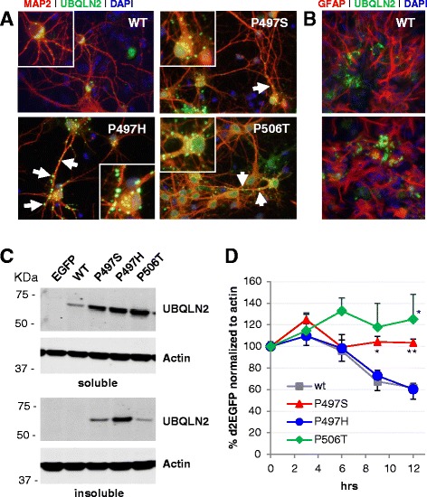
Background: UBQLN2 mutations have recently been associated with familial forms of amyotrophic lateral sclerosis (ALS) and ALS-dementia. UBQLN2 encodes for ubiquilin-2, a member of the ubiquitin-like protein family which facilitates delivery of ubiquitinated proteins to the proteasome for degradation. To study the potential role of ubiquilin-2 in ALS, we used recombinant adeno-associated viral (rAAV) vectors to express UBQLN2 and three of the identified ALS-linked mutants (P497H, P497S, and P506T) in primary neuroglial cultures and in developing neonatal mouse brains.

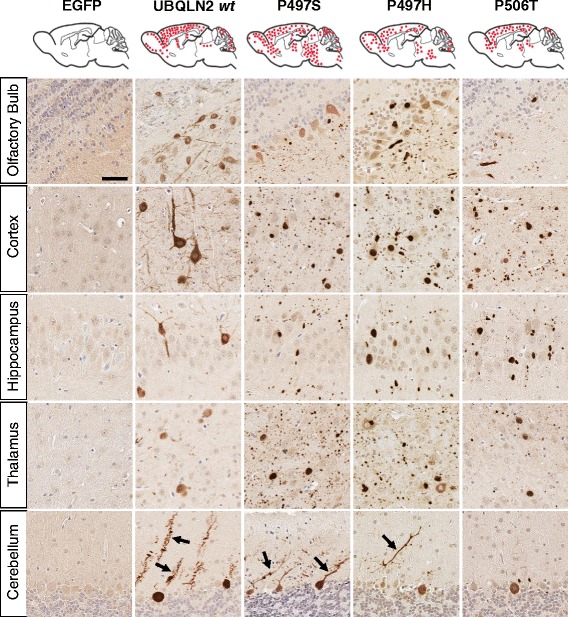
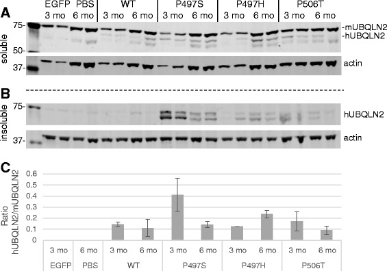
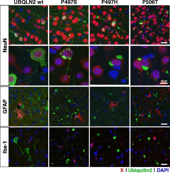
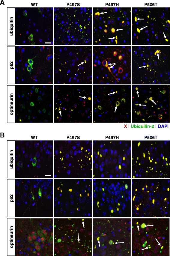
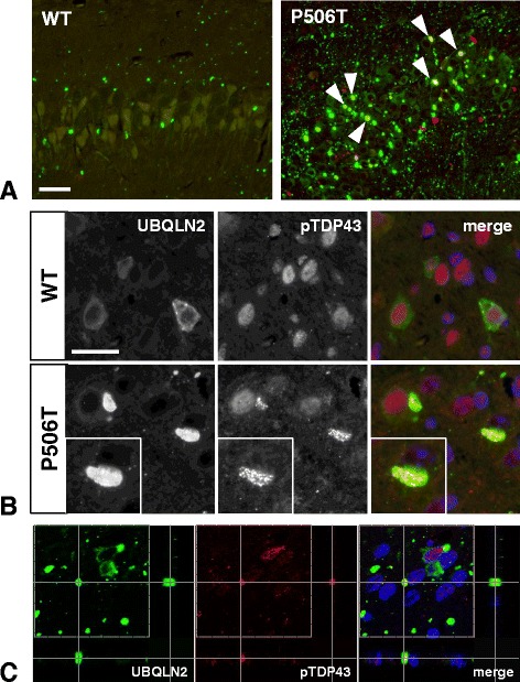
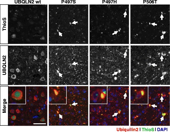
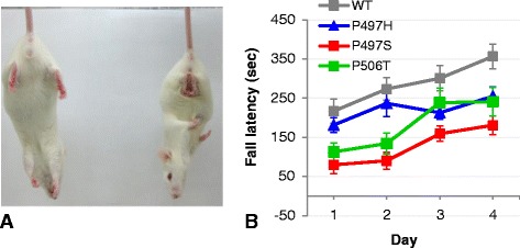
Results: In primary cultures rAAV2/8-mediated expression of UBQLN2 mutants resulted in inclusion bodies and insoluble aggregates. Intracerebroventricular injection of FVB mice at post-natal day 0 with rAAV2/8 expressing wild type or mutant UBQLN2 resulted in widespread, sustained expression of ubiquilin-2 in brain. In contrast to wild type, mutant UBQLN2 expression induced significant pathology with large neuronal, cytoplasmic inclusions and ubiquilin-2-positive aggregates in surrounding neuropil. Ubiquilin-2 inclusions co-localized with ubiquitin, p62/SQSTM, optineurin, and occasionally TDP-43, but were negative for α-synuclein, neurofilament, tau, and FUS. Mutant UBLQN2 expression also resulted in Thioflavin-S-positive inclusions/aggregates. Mice expressing mutant forms of UBQLN2 variably developed a motor phenotype at 3-4 months, including nonspecific clasping and rotarod deficits.

Conclusions: These findings demonstrate that UBQLN2 mutants (P497H, P497S, and P506T) induce proteinopathy and cause behavioral deficits, supporting a "toxic" gain-of-function, which may contribute to ALS pathology. These data establish also that our rAAV model can be used to rapidly assess the pathological consequences of various UBQLN2 mutations and provides an agile system to further interrogate the molecular mechanisms of ubiquilins in neurodegeneration.


Figures

Fig. 1
Fig. 1
Mutant Ubiquilin-2 overexpression results in punctate intracellular accumulations in primary mixed neuroglia cultures. a. Cells transduced with AAV-UBQLN2(WT) show ubiquilin-2 immunoreactivity (green) diffusely present in the cytoplasm and cell processes with few small punctate accumulations. In contrast, UBQLN2 mutants (P497S, P497H, and P506T) result in large intracellular ubiquilin-2 accumulations both in neuronal soma and processes (red, labelled with MAP2). Ubiquilin-2 accumulations in processes have a “bead on a string”-like appearance particularly for P497H and P506T mutants. b. Some ubiquilin-2 accumulations (green) are outside of neurons and colocalized with astrocytes in culture, labeled with GFAP-imunoreactivity (red). c. Western blot of TX-soluble and insoluble fractions show that all UBQLN2 mutants and not WT accumulate in the TX-insoluble/SDS fraction, suggesting formation insoluble aggregates. d. Graph of d2EGFP signal normalized to actin in HEK293 cells transfected with WT and mutant ubiquilin-2. Both P497S and P506T mutants show impaired proteasomal degradation of the d2EGFP reporter compared to the P497H mutant and WT ubiquilin-2. *p < 0.05, **p < 0.01
Fig. 2
Fig. 2
Viral expression of ubiquilin-2 at 6 months. Representative schema of sagittal section shows the overall distribution of AAV-UBQLN2 expression in mouse brain after ICV injection (SBT model). Photos show ubiquilin-2 immunostaining in representative sections animals injected with AAV expressing either EGFP control or WT vs P497S, P497H, or P506T mutant ubiquilin-2. WT ubiqulin-2 expression is homogeneous throughout the neuronal perykaria, including processes. Mutant ubiquilin-2 shows altered subcellular expression, often concentrating in the nucleus, but also resulting cytoplasmic inclusions. Ubiquilin-2-positive “aggregates” are seen also in adjacent neuropil. Arrows point out alteration in purkinje cell dendritic arbors for mutant vs WT ubiquilin-2. (Scale same for all photomicrographs, bar = 50 μm)
Fig. 3
Fig. 3
Viral expression of human ubiquilin-2 in whole brain lysates. Western blots of brain lysates from 3 and 6 month animals demonstrate sustained expression of WT ubiquilin-2 and mutants. In the Trition-X100 soluble fraction (a) three bands are seen for ubiquilin-2: top is mouse UBQLN2 whereas middle and lower (truncated?) bands represent human UBQLN2. b) Only mutant forms of human UBQLN2 are seen in the Triton insoluble fractions. c) Graph of human vs endogenous mouse UBQLN2 expression in whole brain (Triton soluble) lysates. N = 2–3 sample each with mean ± SD ratio shown
Fig. 4
Fig. 4
AAV expression of UBQLN2 WT and mutants is specific to neurons. Images are merged photos of representative cortical areas from 6 month mice stained with immunofluorescence for NeuN/GFAP/Iba-1 (red), ubiquilin-2 (green), and DAPI (blue). The top 2 rows show colocalization of ubiquilin-2 accumulations with NeuN-positive neurons. Row 2 includes high-power confocal images that demonstrate differences in the distribution of ubiquilin-2-containing inclusions in NeuN labeled neurons; large inclusions are seen for all mutant forms in contrast to WT ubiquilin-2. Glial markers GFAP (row 3) and Iba-1 (row 4) rarely colocalize with ubiquilin-2. (Scale bar = 50 μm unless otherwise noted)
Fig. 5
Fig. 5
Ubiquilin-2 inclusions colocalize with ubiquitin, p62, and optineurin. Merged immunofluorescent images are from a) cortex and b) hippocampus from 6 month mice and show staining for ubiquitin/p62/optineurin (red), ubiquilin-2 (green), and DAPI (blue). In contrast to WT, pathological (mutant) ubiquilin-2 form large intracellular and neuropil inclusions that frequently colocalize (indicated as yellow, representing overlap red and green signal) with ubiquitin, p62, and optineurin. (bar = 25 μm)
Fig. 6
Fig. 6
TDP43 colocalizes with ubiquilin-2 in mice expressing mutant UBQLN2 (P506T). a) Low power merged immunofluorescent images of CA3 hippocampus from mice injected with rAAV expressing WT or P506T mutant ubiquilin-2 and aged 6 months. TDP43 (red) colocalizes (arrowheads) with several ubiquilin-2-positive (green) inclusions in mutant P506T expressing mice. (bar = 25 μm) b) Higher power photomicrographs show cytoplasmic TDP43 puncta stained with the phospho-TDP43 antibody (403–404) within a large cytoplasmic ubiquilin-2-positive inclusion. (bar = 25 μm) c) Confocal Z-slice section analysis of ubiquilin-2 inclusions (green) similarly demonstrates colocalization of phosphorylated TDP43 (red) in brain tissue from mice expressing mutant UBQLN2 (P506T)
Fig. 7
Fig. 7
Mutant ubiquilin-2 expression induces ThioS-positive inclusions. Photos show Thioflavin-S staining that colocalizes (arrows; yellow in merged images) with intracellular ubiquilin-2-positive inclusions seen in mice injected with rAAV expressing mutant but not WT ubiquilin-2. Images shown are from mice aged 6 months, but similar findings were seen also in younger mice. (bar = 50 μm)
Fig. 8
Fig. 8
Behavioral deficits in mice. a) Mice expressing ALS-mutant ubiquilin-2 develop a clasping phenotype at 3–4 months. b) Rotarod performance for mice at 3 months expressing mutant ubiquilin-2 P497S (p < 0.0001) and P506T (p < 0.01) was significantly impaired compared to those expressing WT ubiquilin-2. Data shown as mean ± SEM; N = 9 for each group

References

    1. Deng HX, Chen W, Hong ST, Boycott KM, Gorrie GH, Siddique N, et al. Mutations in UBQLN2 cause dominant X-linked juvenile and adult-onset ALS and ALS/dementia. Nature. 2011;477:211–5. doi: 10.1038/nature10353. - DOI - PMC - PubMed
    1. Synofzik M, Maetzler W, Grehl T, Prudlo J, Vom Hagen JM, Haack T, et al. Screening in ALS and FTD patients reveals 3 novel UBQLN2 mutations outside the PXX domain and a pure FTD phenotype. Neurobiol Aging. 2012;33:2949 e2913–2947. doi: 10.1016/j.neurobiolaging.2012.07.002. - DOI - PubMed
    1. Fahed AC, McDonough B, Gouvion CM, Newell KL, Dure LS, Bebin M, Bick AG, Seidman JG, Harter DH, Seidman CE: UBQLN2 mutation causing heterogeneous X-linked dominant neurodegeneration. Annals of neurology 2014, 75(5):793-798. - PMC - PubMed
    1. Marin I. The ubiquilin gene family: evolutionary patterns and functional insights. BMC Evol Biol. 2014;14:63. doi: 10.1186/1471-2148-14-63. - DOI - PMC - PubMed
    1. Rothenberg C, Srinivasan D, Mah L, Kaushik S, Peterhoff CM, Ugolino J, et al. Ubiquilin functions in autophagy and is degraded by chaperone-mediated autophagy. Hum Mol Genet. 2010;19:3219–32. doi: 10.1093/hmg/ddq231. - DOI - PMC - PubMed

Publication types

MeSH terms