. 2015 Jul 7;6(4):e00379.
doi: 10.1128/mBio.00379-15.

Chemorepulsion from the Quorum Signal Autoinducer-2 Promotes Helicobacter pylori Biofilm Dispersal

Affiliations

Chemorepulsion from the Quorum Signal Autoinducer-2 Promotes Helicobacter pylori Biofilm Dispersal

Jeneva K Anderson et al. mBio. .

Abstract

The gastric pathogen Helicobacter pylori forms biofilms on abiotic and biotic surfaces. We have shown previously that H. pylori perceives the quorum signal autoinducer-2 (AI-2) as a chemorepellent. We report here that H. pylori chemorepulsion from endogenous AI-2 influences the proportions and spatial organization of cells within biofilms. Strains that fail to produce AI-2 (∆luxS strains) or are defective for chemotaxis (∆cheA strains) formed more spatially homogenous biofilms with a greater proportion of adherent versus planktonic cells than wild-type biofilms. Reciprocally, a strain that overproduced AI-2 (luxS(OP)) formed biofilms with proportionally fewer adherent cells. Along with the known AI-2 chemoreceptor, TlpB, we identified AibA and AibB, two novel periplasmic binding proteins that are required for the AI-2 chemorepulsion response. Disruptions in any of the proteins required for AI-2 chemotaxis recapitulated the biofilm adherence and spatial organization phenotype of the ∆luxS mutant. Furthermore, exogenous administration of AI-2 was sufficient to decrease the proportion of adherent cells in biofilms and promote dispersal of cells from biofilms in a chemotaxis-dependent manner. Finally, we found that disruption of AI-2 production or AI-2 chemotaxis resulted in increased clustering of cells in microcolonies on cultured epithelial cells. We conclude that chemotaxis from AI-2 is a determinant of H. pylori biofilm spatial organization and dispersal.

Importance: Bacterial biofilms are ubiquitous in nature, but the mechanisms governing their assembly and spatial organization are not fully understood. Bacterial communication through quorum sensing has been shown to influence biofilm growth through the regulation of biofilm genes. Our study revealed a new role for quorum sensing in biofilms through rapid chemotactic responses to quorum signals. Specifically, we studied how chemorepulsion of Helicobacter pylori from the universal quorum signal autoinducer-2 (AI-2) shapes the spatial organization of its biofilms. We demonstrate that the chemorepulsive response of H. pylori to AI-2 is necessary to promote its dispersal from biofilms grown on both abiotic and biotic surfaces and is sufficient to promote dispersal in a chemotaxis-dependent manner. This work has broad implications for understanding the mechanisms by which endogenously produced microbial compounds shape the assembly and spatial organization of microbial communities in their environments.


Figures

FIG 1
FIG 1
Endogenous AI-2 production and chemotaxis alter biofilm organization. (A to E) Epifluorescence images of wild-type and mutant G27 H. pylori biofilms grown on glass slides for 5 days. Cells were fixed and stained with DAPI (white). Bar, 100 µm. (F) Lacunarity scores for biofilm images. Each dot represents the lacunarity score for a single epifluorescence biofilm image postthresholding, with corresponding means and standard deviations. (G) Schematic of glass frit biofilm assay with crystal violet analysis and examples of percentages of cells in the biofilm calculation. (H) Effect of AI-2 production and chemotaxis on adherence of cells in biofilms on glass frit after 2 days, as quantified by the crystal violet assay. Error bars represent standard errors of the means of the results of a minimum of three experiments for each strain. Daggers indicate a significant (P < 0.05) difference from the wild-type results determined using Student’s t test (F) or one-way analysis of variance (H).
FIG 2
FIG 2
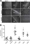
AI-2 chemotaxis requires two periplasmic binding proteins, AibA and AibB, that bind AI-2 independently. (A) Chemotaxis response of wild-type and ΔHPG27_277aibA) and ΔHPG27_431aibB) mutant H. pylori to brucella broth (BB10), 100 mM HCl, and 100 µM synthetic DPD (AI-2). Representative wet-mount images of bacterial cells (white dots) are shown. Formation of a barrier (marked by white arrows) indicates a chemotactic response. Bar, 200 µm. (B) In vitro AI-2 binding assay performed with purified proteins using a V. harveyi bioluminescence readout. Percentages of relative luminescence units (RLU) were normalized to an AI-2 positive control in each independent experiment. Error bars represent standard errors of the means of the results of comparisons of experimental data. Daggers indicate a significant (P < 0.05) difference from the buffer negative-control results determined using Student’s t test.
FIG 3
FIG 3
Chemorepulsion from AI-2 is necessary for biofilm organization. (A to G) Epifluorescence images of AI-2 chemotaxis-defective mutants and complemented-strain biofilms grown on glass slides for 5 days. Cells were fixed and stained with DAPI (white). (H) Lacunarity scores for biofilm images. Each point represents the lacunarity score for a single epifluorescence biofilm image postthresholding, with corresponding means and standard deviations. (I) Effect of AI-2 chemotaxis disruption on adherence of cells in biofilms on glass frit after 2 days, as quantified by the crystal violet assay. Error bars represent standard errors of the means of the results of a minimum of 3 experiments for each strain. Daggers indicate a significant (P < 0.05) difference from the wild-type results determined using Student’s t test (H) or one-way analysis of variance (I).
FIG 4
FIG 4
Chemorepulsion from AI-2 is sufficient to decrease adherence in biofilms. (A) Change in adherence of strains in biofilm with addition of exogenous AI-2 (37 nM) at the time of inoculation. Daggers indicate a significant (P < 0.05) difference from 0 (no change) determined using Student’s t test. (B) Fraction changes in biofilm size (grey bars) and numbers of planktonic cells (white bars) measured 72 h after the initiation of the biofilms and 24 h after rinsing and exchange with fresh media containing 37 nM exogenous AI-2. Daggers indicate a significant (P < 0.05) difference from a value of 1 (no change) determined using Student’s t test. Box plots show means and standard deviations of the results of a minimum of three experiments for each strain.
FIG 5
FIG 5
Disruption of either AI-2 production or AI-2 chemotaxis results in larger microcolonies on cultured epithelial cell monolayers. (A to C) Representative images of WT, ΔluxS, and ΔtlpB H. pylori microcolonies on polarized MDCK monolayers at 3 days postinfection. H. pylori cells are visualized in red, nuclei in blue, and cell junctions in green. Bar, 10 µm. (D) Estimated numbers of attached bacteria on the MDCK epithelial cell layer at 5 min and 3 days postinfection for all strains. Each point represents the estimated number of bacteria averaged over multiple images per transwell. Error bars represent standard errors of the means. Daggers represent significant increases in numbers of attached bacteria from 5 min to 3 days of attachment for each strain. (E) Plot of the surface area covered by contiguous clusters of bacterial cells (microcolonies) for each strain at 5 min (predominantly isolated single cells) and 3 days postinoculation. Each dot represents the average area (in pixels) of all the microcolonies in an image. Three images were taken per transwell, with three replicate transwells per experiment. Error bars represent standard errors of the means. Daggers represent significant (P < 0.05) increases of pixel sizes compared to the WT results through the use of one-way analysis of variance.

References

    1. Wozniak DJ, Parsek MR. 2014. Surface-associated microbes continue to surprise us in their sophisticated strategies for assembling biofilm communities. F1000PrimeRep 6:26. doi:10.12703/P6-26. - DOI - PMC - PubMed
    1. De la Fuente-Núñez C, Reffuveille F, Fernández L, Hancock RE. 2013. Bacterial biofilm development as a multicellular adaptation: antibiotic resistance and new therapeutic strategies. Curr Opin Microbiol 16:580–589. doi:10.1016/j.mib.2013.06.013. - DOI - PubMed
    1. Elias S, Banin E. 2012. Multi-species biofilms: living with friendly neighbors. FEMS Microbiol Rev 36:990–1004. doi:10.1111/j.1574-6976.2012.00325.x. - DOI - PubMed
    1. Donlan RM. 2002. Biofilms: microbial life on surfaces. Emerg Infect Dis 8:881–890. doi:10.3201/eid0809.020063. - DOI - PMC - PubMed
    1. Davey ME, George AO. 2000. Microbial biofilms: from ecology to molecular genetics. Microbiol Mol Biol Rev 64:847–867. doi:10.1128/MMBR.64.4.847-867.2000. - DOI - PMC - PubMed

Publication types

MeSH terms

LinkOut - more resources