. 2015 Jul 7;6(4):e00759.
doi: 10.1128/mBio.00759-15.

NS5A Domain 1 and Polyprotein Cleavage Kinetics Are Critical for Induction of Double-Membrane Vesicles Associated with Hepatitis C Virus Replication

Affiliations

NS5A Domain 1 and Polyprotein Cleavage Kinetics Are Critical for Induction of Double-Membrane Vesicles Associated with Hepatitis C Virus Replication

Inés Romero-Brey et al. mBio. .

Abstract

Induction of membrane rearrangements in the cytoplasm of infected cells is a hallmark of positive-strand RNA viruses. These altered membranes serve as scaffolds for the assembly of viral replication factories (RFs). We have recently shown that hepatitis C virus (HCV) infection induces endoplasmic reticulum-derived double-membrane vesicles (DMVs) representing the major constituent of the RF within the infected cell. RF formation requires the concerted action of nonstructural action of nonstructural protein (NS)3, -4A, protein (NS)3 -4A, -4B, -5A, and -5B. Although the sole expression of NS5A is sufficient to induce DMV formation, its efficiency is very low. In this study, we dissected the determinants within NS5A responsible for DMV formation and found that RNA-binding domain 1 (D1) and the amino-terminal membrane anchor are indispensable for this process. In contrast, deletion of NS5A D2 or D3 did not affect DMV formation but disrupted RNA replication and virus assembly, respectively. To identify cis- and trans-acting factors of DMV formation, we established a trans cleavage assay. We found that induction of DMVs requires full-length NS3, whereas a helicase-lacking mutant was unable to trigger DMV formation in spite of efficient polyprotein cleavage. Importantly, a mutation accelerating cleavage kinetics at the NS4B-5A site diminished DMV formation, while the insertion of an internal ribosome entry site mimicking constitutive cleavage at this boundary completely abolished this process. These results identify key determinants governing the biogenesis of the HCV RF with possible implications for our understanding of how RFs are formed in other positive-strand RNA viruses.

Importance: Like all positive-strand RNA viruses, hepatitis C virus (HCV) extensively reorganizes intracellular membranes to allow efficient RNA replication. Double-membrane vesicles (DMVs) that putatively represent sites of HCV RNA amplification are induced by the concerted action of viral and cellular factors. However, the contribution of individual proteins to this process remains poorly understood. Here we identify determinants in the HCV replicase that are required for DMV biogenesis. Major contributors to this process are domain 1 of nonstructural protein 5A and the helicase domain of nonstructural protein 3. In addition, efficient DMV induction depends on cis cleavage of the viral polyprotein, as well as tightly regulated cleavage kinetics. These results identify key determinants governing the biogenesis of the HCV replication factory with possible implications for our understanding of how this central compartment is formed in other positive-strand RNA viruses.


Figures

FIG 1
FIG 1
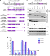
Deletions within NS5A and their impact on RNA replication and polyprotein cleavage. (A) Schematic representation of the subgenomic replicons used in this study. The 3′ UTR of HCV and the IRES of encephalomyocarditis virus downstream of the firefly luciferase gene (Luc; gray box) are shown as stem-loop structures. An enlarged view of the organization of the NS5A domain is shown below. The numbers at the top refer to amino acid residues of the JFH-1 polyprotein, and numbers in parentheses refer to residues of NS5A. Deletion mutants are shown below; the names of the mutants are shown to the left of the illustrations. The numbers in parentheses refer to the amino acid (aa) residues of the polyprotein that were removed. (B) Replication kinetics of NS5A deletion mutants. Huh7.5 cells were transfected with subgenomic Luc reporter replicons. Cells were harvested 4, 24, 48, and 72 h post electroporation (hpe), and the luciferase activity in the lysates was determined. Values were normalized to the 4-h value, reflecting transfection efficiency. A replication-deficient mutant encoding an inactive NS5B polymerase served as a negative control (ΔGDD). RLU, relative light units; WT, wild type; LOD, limit of detection. (C) Polyprotein processing of NS5A mutants. The basic construct used for expression of the NS3-5B polyprotein and NS5A mutant proteins derived therefrom is shown on the top. It contains the promoter (Pm) of the T7 RNA polymerase (gray box) and the IRES of encephalomyocarditis virus upstream of the polyprotein coding region. Huh7-Lunet/T7 cells were transfected with expression constructs specified above the lanes, and 24-h later, cells were lysed and HCV proteins were detected by Western blotting with various NS5A-, NS4B-, and NS3-specific antibodies as specified on the right of each panel. β-Actin served as a loading control. The values on the left are the molecular masses of the standards used. The uncleaved NS4B-ΔD15A precursor detected in the NS5A (9E10)- and NS4B-specific immunoblot analysis is indicated by arrowheads. The two phosphorylated variants of wild-type NS5A in the upper panel are labeled with asterisks, and they are referred to as p56 and p58.
FIG 2
FIG 2
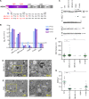
Deletions within NS5A and their impact on the formation of DMVs. (A to G) Cells were transfected with the expression constructs shown to the left of the panels, and 24 h later, cells were fixed and processed for EM by cryofixation and epon embedding. (H, I) Quantitative analyses of DMV diameters (H) and numbers of DMVs per square micrometer of cell surface area (I). Fifteen cell profiles from two independent experiments (10 and 5 cell profiles, respectively) were analyzed. The P values shown were calculated by using unpaired Student t tests; n.s., nonsignificant; *, P < 0.01; **, P < 0.001; ***, P < 0.0001; n.d., not determined because of absence of DMVs; WT, wild type. Green horizontal lines represent mean values.
FIG 3
FIG 3
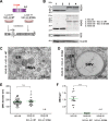
Essential role of NS5A D1 in DMV formation. (A) Schematic of the NS3-5B polyprotein expression construct lacking NS5A D1 and containing a GFP insertion in D3 (pTM_NS3-5B_ΔD1-GFP). Note that this GFP insertion does not affect the functionality of NS5A (24). Pm, promoter. (B) Abundance of variant NS5A, NS4B, and NS3 proteins in Huh7-Lunet/T7 cells transfected with pTM_NS3-5B_ΔD1-GFP in comparison with that in cells transfected with pTM_NS3-5B_WT. Cell lysates prepared 24 h after transfection were analyzed by Western blotting with monospecific antibodies. The uncleaved NS4B-5A precursor is indicated by an arrowhead. WT, wild type. (C) Twenty-four hours after transfection, Huh7-Lunet/T7 cells were analyzed by fluorescence microscopy to allocate GFP-positive cells grown on patterned sapphire discs. (D) Superposition of a low-magnification electron micrograph and the fluorescence image of the cell boxed in panel C. (E and F) Higher-magnification micrographs of the boxed area of this cell, revealing high numbers of LDs but an absence of DMVs.
FIG 4
FIG 4
Determinants within the N-terminal AH and D1 of NS5A required for DMV formation. (A) Schematic representation of the domain organization of NS5A and the positions of the mutations introduced. The amino acid (aa) sequence of the N-terminal AH is shown at the bottom, with the amino acid residues that were replaced in red. (B) Replication kinetics of NS5A mutants as determined with subgenomic luciferase reporter replicons. For further details, see the legend to Fig. 1. RLU, relative light units; hpe, hours post electroporation; LOD, limit of detection. (C) Abundance of HCV proteins in Huh7-Lunet/T7 cells transfected with NS3-5B polyprotein expression constructs. Cell lysates prepared 24 h after transfection were analyzed by Western blotting with monospecific antibodies. The uncleaved NS4B-5A precursor is indicated by an arrowhead. WT, wild type. (D and E) Cells transfected in the same way were grown on sapphire discs and processed for EM as described in the legend to Fig. 2. Representative images of the mutants specified to the left of the panels are shown. Magnified views of the boxed areas are shown to the right. MVB, multivesicular body. (F and G) Quantitative analysis of EM images. For further details, see the legend to Fig. 2. The data presented for the wild type are the same as those shown in Fig. 2H and I because the mutant sets shown in Fig. 2 and 4 were analyzed in parallel, along with the wild type. n.d., not determined; n.s., nonsignificant; **, P < 0.001; ***, P < 0.0001.
FIG 5
FIG 5
Formation of DMVs requires the NS3 helicase domain. (A) Schematic drawing of polyprotein expression constructs. Numbers refer to the JFH-1 polyprotein; For further details, see the legend to Fig. 1A. Pm, promoter. (B) Western blot analysis of lysates of cells harvested 24 h after transfection. β-Actin served as a loading control. WT, wild type. (C and D) Analyses of transfected cells by EM. Cells grown on sapphire discs were transfected with the constructs shown to the left of the panels, and 24 h later, cells were processed for EM as described in the legend to Fig. 2. Note that only SMVs were found in the helicase deletion mutant (30 cell profiles from two independent experiments analyzed).
FIG 6
FIG 6
Efficiency of DMV formation is determined by polyprotein cleavage in cis. (A) Schematic of the experimental approach used for trans cleavage of the polyprotein. Huh7-Lunet/T7-derived cell lines stably expressing wild-type (WT) NS3-4A or an inactive protease (S139A) were transfected with the NS4B-5B polyprotein expression construct. (B) Western blot analysis of cell lysates prepared 24 h after transfection. The constructs and cell lines used are specified on the right. β-Actin served as a loading control. (C and D) Representative EM images of cells expressing the NS4B-5B polyprotein and wild-type NS3-4A or an inactive NS3 protease mutant. (E and F) Quantitative analyses of DMV diameters (E) and numbers of DMVs per square micrometer of cell surface area. (F) From two independent experiments, a total of 90 and 30 DMVs and 10 and 5 cell profiles were analyzed for NS3-5B- and NS4B-5B + NS3-4A WT-expressing cells, respectively. n.d., not determined; n.s., nonsignificant; ***, P < 0.0001.
FIG 7
FIG 7
Enhancement of cleavage kinetics at the NS4B-5A site negatively affects RNA replication and DMV formation. (A) The basic polyprotein expression construct is depicted at the top (cf. Fig. 1A). The introduced double mutation reported to accelerate cleavage kinetics at the NS4B-5A site (20) is shown at the bottom (construct P43VV fast). Huh7-Lunet/T7 cells were transfected with each of these constructs and 1 day later incubated with [35S]methionine/cysteine-containing medium for 1 h. The medium was removed, and cells were harvested (0 min) or incubated in nonradioactive medium for 10 or 20 min. NS5A-containing proteins were isolated by NS5A-specific immunoprecipitation and analyzed by SDS-PAGE and autoradiography. HCV proteins are specified on the right, and the positions of molecular weight marker proteins are shown on the left. Pm, promoter; WT, wild type. (B) Replication kinetics of subgenomic luciferase reporter replicons containing cleavage site mutations. The wild type and the NS5B polymerase-dead mutant (ΔGDD) served as positive and negative controls, respectively. LOD, limit of detection. For further details, see the legend to Fig. 1B. Luc, luciferase; RLU, relative light units; hpe, hours postel ectroporation. (C) Representative EM images of cells expressing the NS3-5B polyprotein containing the cleavage site mutation. The right panel is an enlarged view of the boxed region in the left panel. (D and E) Quantitative analyses of DMV diameters (D) and numbers of DMVs per cell surface area (E). Ten cell profiles from two independent experiments were analyzed.
FIG 8
FIG 8
Uncleaved NS4B-5A is required for RNA replication and DMV formation. (A) The design of the subgenomic luciferase reporter replicon containing the IRES of encephalomyocarditis virus between NS4B and NS5A is shown on the top. Replication kinetics, as determined by transient transfection of Huh7.5 cells, are displayed below. Values were normalized to the 4-h values, reflecting transfection efficiency. For further details, see the legend to Fig. 1B. Luc, luciferase; hpe, hours post electroporation; RLU, relative light units; LOD, limit of detection; WT, wild type. (B) Schematic representation of the expression construct transfected into Huh7-Lunet/T7 cells. Twenty-four hours after transfection, cells were lysed and the abundance of HCV NS proteins was determined by Western blotting. Calnexin served as a loading control. (C) In parallel, a fraction of the cells was fixed and analyzed by EM as described in the legend to Fig. 2. Note that only SMVs were detected in cells transfected with the IRES insertion mutant. A total of 30 cell profiles obtained from two independent experiments were analyzed. The image on the right is a higher-magnification view of the area boxed on the left.

References

    1. Romero-Brey I, Bartenschlager R. 2014. Membranous replication factories induced by plus-strand RNA viruses. Viruses 6:2826–2857. doi:10.3390/v6072826. - DOI - PMC - PubMed
    1. Paul D, Bartenschlager R. 2013. Architecture and biogenesis of plus-strand RNA virus replication factories. World J Virol 2:32–48. doi:10.5501/wjv.v2.i2.32. - DOI - PMC - PubMed
    1. Ferraris P, Beaumont E, Uzbekov R, Brand D, Gaillard J, Blanchard E, Roingeard P. 2013. Sequential biogenesis of host cell membrane rearrangements induced by hepatitis C virus infection. Cell Mol Life Sci 70:1297–1306. doi:10.1007/s00018-012-1213-0. - DOI - PMC - PubMed
    1. Romero-Brey I, Merz A, Chiramel A, Lee JY, Chlanda P, Haselman U, Santarella-Mellwig R, Habermann A, Hoppe S, Kallis S, Walther P, Antony C, Krijnse-Locker J, Bartenschlager R. 2012. Three-dimensional architecture and biogenesis of membrane structures associated with hepatitis C virus replication. PLoS Pathog 8:e1003056. doi:10.1371/journal.ppat.1003056. - DOI - PMC - PubMed
    1. Egger D, Wölk B, Gosert R, Bianchi L, Blum HE, Moradpour D, Bienz K. 2002. Expression of hepatitis C virus proteins induces distinct membrane alterations including a candidate viral replication complex. J Virol 76:5974–5984. doi:10.1128/JVI.76.12.5974-5984.2002. - DOI - PMC - PubMed

Publication types

LinkOut - more resources