. 2015 Jul 8;11(7):e1005028.
doi: 10.1371/journal.ppat.1005028. eCollection 2015 Jul.

Neuronal Interferon Signaling Is Required for Protection against Herpes Simplex Virus Replication and Pathogenesis

Affiliations

Neuronal Interferon Signaling Is Required for Protection against Herpes Simplex Virus Replication and Pathogenesis

Pamela C Rosato et al. PLoS Pathog. .

Abstract

Interferon (IFN) responses are critical for controlling herpes simplex virus 1 (HSV-1). The importance of neuronal IFN signaling in controlling acute and latent HSV-1 infection remains unclear. Compartmentalized neuron cultures revealed that mature sensory neurons respond to IFNβ at both the axon and cell body through distinct mechanisms, resulting in control of HSV-1. Mice specifically lacking neural IFN signaling succumbed rapidly to HSV-1 corneal infection, demonstrating that IFN responses of the immune system and non-neuronal tissues are insufficient to confer survival following virus challenge. Furthermore, neurovirulence was restored to an HSV strain lacking the IFN-modulating gene, γ34.5, despite its expected attenuation in peripheral tissues. These studies define a crucial role for neuronal IFN signaling for protection against HSV-1 pathogenesis and replication, and they provide a novel framework to enhance our understanding of the interface between host innate immunity and neurotropic pathogens.


Conflict of interest statement

The authors have declared that no competing interests exist.

Figures

Fig 1
Fig 1. Paracrine IFNβ signaling at the cell body and distal axon reduces HSV-1 titers upon axonal infection.
A) Diagram of Campenot chambers that were modified by removing one of two central barriers. Trigeminal ganglia (TG) neurons from adult mice were isolated and seeded in soma compartment. Micrograph shows βIII-tubulin staining of neurons extending axons through a single barrier guided by grooves to the axonal compartment. Scale bar = 250μm. B) Diagram depicting the compartment where virus was added (indicated by a red virion), and where sampling occurred (blue background). Titers of WT (strain 17) or Δγ34.5 viruses in the soma compartment 72 hours post-axonal infection with 108 PFU in 129SVEV and STAT1-/- neurons. Cultures were untreated or treated with 12.5U/mL IFNβ in the soma or axon compartment. Dashed line represents the limit of detection. Error bars represent SEM of a minimum of 3 experiments with >2 chambers each. Significance was evaluated by two-way ANOVA where ** p<0.01, ***p<0.001.
Fig 2
Fig 2. Axonal IFNβ signaling restricts HSV-1 titers through mechanisms independent of establishment of an antiviral state at the soma.
A) Titers of WT (strain 17) or Δγ34.5 viruses in the soma compartment of 129SVEV neuron cultures 72 hours post infection via the soma. Cultures were untreated (white bars) or treated with 12.5U/mL IFNβ in the soma (grey bars) or axon compartment (black bars) 18 hours prior to infection. B) Titers of VSV in the soma compartment 24 hours post infection via the soma of 129SVEV neuron cultures. Cultures were treated with IFNβ as in (A). C) Representative image of immunofluorescence staining for STAT1 (red) in chambers treated with IFNβ at the soma for 3 hours (left) or axon for 3, 5, 7 or 16 hours (right). Cells were counterstained for nuclei (DAPI, blue). DiO (green) was added to the axon compartment, thereby labeling neurons that extended axons through the central barrier. Scale bar = 50μm. D) Expression of IFIT1 and ISG15 transcripts in the soma compartment of chambers treated with IFNβ for 6 or 12 hours at the soma or axon. Error bars represent SEM of a minimum of 4 samples over 2 experiments. E) HSV genome copy number of WT (strain 17) or Δγ34.5 virus measured from the soma compartment of axonally infected 129SVEV neuron cultures at 1hpi and 3hpi. Cultures were treated with 100μM ACV, and with 12.5 U/mL IFNβ in the axon compartment for 18 hours, or 10μM capsaicin (red bar) in both compartments for 0.5 hour prior to infection with 108 PFU. Error bars represent SEM of a minimum of 3 repeats with >3 chambers each. Two-way ANOVA was performed where *p<0.05, ** p<0.01, ***p<0.001.
Fig 3
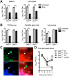
Fig 3. Validation of Stat1N-/- mouse.
Titers of VSV in BMDCs or fibroblasts (A) or TG neurons, satellite glial cells, or astrocytes (B) isolated from Stat1N-/- or Stat1fl/fl mice. Cells were untreated or treated with 12.5 units/mL IFNβ and titers were measured at 24hpi. Error bars represent SEM of at least two experiments with ≥2 samples each. C) Immunofluorescence staining of brain tissue from 3 week old progeny from a cross of TdTomato reporter and Nestin Cre mice. TdTomato fluorescence indicates cells that express or expressed CRE recombinase. White arrow marks Iba1+ microglia (green). Cells were counterstained for DAPI (blue). Scale bar = 20μm. D) Viral titers from corneal eye swabs of Stat1N-/- or Stat1fl/fl mice infected via corneal scarification with 2 x 10⁶ PFU/eye WT (stain 17) or Δγ34.5 virus. Dashed line represents the limit of detection, and error bars represent SEM of a minimum of 7 mice, over at least 2 experiments. Two-way ANOVA with Bonferroni correction was performed where one symbol indicates p<0.05, and three symbols indicate p<0.001. Unless noted with brackets, * indicates significant differences between Stat1N-/- WT and Stat1N-/- Δγ34.5; and ‡ between Stat1fl/fl WT and Stat1fl/fl Δγ34.5.
Fig 4
Fig 4. Neural STAT1 expression is critical for controlling HSV-1 replication in vivo.
Viral titers in the TG, brain stem and brain of Stat1N-/- or Stat1fl/fl mice infected via corneal scarification with 2 x 10⁶ PFU/eye WT (strain 17) or Δγ34.5 virus. Viral titers were measured at 3 dpi (A) and 5 dpi (B). Dashed lines represent the limit of detection. Error bars represent SEM of a minimum of 13 mice total over 2 experiments. Two-way ANOVA was performed where ** p<0.01, ***p<0.001.
Fig 5
Fig 5. Neural deletion of STAT1 affects viral tropism in the trigeminal ganglia.
A) Immunofluorescence of TG sections from Stat1N-/- or Stat1fl/fl mice 5 days post infection with 2 x 10⁶ PFU/eye WT (strain 17) or Δγ34.5 virus via the cornea. Tissue sections show immunostaining for HSV antigen (red), the neuronal marker NeuN (green), and nuclei (DAPI, blue). The ophthalmic branch of the TG is indicated and white dotted lines mark the trigeminal root entry zone (TREZ) delineating the boundary between PNS and CNS. Scale bar = 500 μm. B) Quantification of the percentage of total cells that are HSV antigen-positive. C) Quantification of the percent of total neurons that are HSV antigen-positive. D) Quantification of the percent of HSV antigen-positive cells that are neurons. Quantification was done using ImageJ/Fuji. Individual data points represent the average of >5 tissue slices per TG with a minimum of 7 TGs over 3 experiments. Student’s t-test was performed where *p<0.05, ** p<0.01, ***p<0.001.
Fig 6
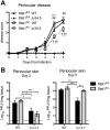
Fig 6. Viral zosteriform spread and pathogenesis in non-neuronal tissues of Stat1N-/- mice.
A) Periocular disease in Stat1N-/- or Stat1fl/fl mice infected via the cornea with 2 x 10⁶ PFU/eye WT (strain 17) or Δγ34.5 virus. Disease scoring was based on a 1–4 scale. B) Titers of periocular skin from Stat1N-/- or Stat1fl/fl mice infected as in (A). Data points represent the average of 2 skin punch titers from each eye. Dashed lines delineate the limit of detection, and error bars represent SEM of a minimum 12 mice, over at least 2 experiments. Two-way ANOVA was performed where one symbol indicates p<0.05, two symbols p<0.01, and three symbols p<0.001. Unless noted with brackets, * indicates significant differences between Stat1N-/- WT and Stat1N-/- Δγ34.5; # between Stat1fl/fl WT and Stat1N-/- Δγ34.5; and † between Stat1fl/fl Δγ34.5 and Stat1N-/- Δγ34.5.
Fig 7
Fig 7. Neural STAT1 expression is required for host survival.
Survival of Stat1N-/- or Stat1fl/fl mice infected via the cornea with 2 x 10⁶ PFU/eye strain 17 (st17) or Δγ34.5 virus. Mice were monitored over time and were euthanized upon reaching end-point criteria, here referred to as survival. Data represent two independent experiments where Stat1fl/fl WT n = 14 mice, Stat1N-/- WT n = 12, Stat1fl/fl Δγ34.5 n = 13 and Stat1N-/- Δγ34.5 n = 12 mice. Survival was plotted on a Kaplan-Meier curve and statistics performed with Log-rank test where ** p<0.01, ***p<0.001.

References

    1. Blyth WA, Harbour DA, Hill TJ. Pathogenesis of zosteriform spread of herpes simplex virus in the mouse. J Gen Virol. 1984;65 (Pt 9): 1477–1486. - PubMed
    1. Smith G. Herpesvirus transport to the nervous system and back again. Annu Rev Microbiol. 2012;66: 153–176. 10.1146/annurev-micro-092611-150051 - DOI - PMC - PubMed
    1. Xu F, Sternberg MR, Kottiri BJ, McQuillan GM, Lee FK, Nahmias AJ, et al. Trends in herpes simplex virus type 1 and type 2 seroprevalence in the United States. JAMA J Am Med Assoc. 2006;296: 964–973. - PubMed
    1. Rowe AM, St Leger AJ, Jeon S, Dhaliwal DK, Knickelbein JE, Hendricks RL. Herpes keratitis. Prog Retin Eye Res. 2013;32: 88–101. 10.1016/j.preteyeres.2012.08.002 - DOI - PMC - PubMed
    1. Whitley RJ, Gnann JW. Viral encephalitis: familiar infections and emerging pathogens. Lancet. 2002;359: 507–513. - PubMed

Publication types

Substances