. 2015 Aug;19(8):2006-18.
doi: 10.1111/jcmm.12581. Epub 2015 Jul 8.

Functional abnormalities in iPSC-derived cardiomyocytes generated from CPVT1 and CPVT2 patients carrying ryanodine or calsequestrin mutations

Affiliations

Functional abnormalities in iPSC-derived cardiomyocytes generated from CPVT1 and CPVT2 patients carrying ryanodine or calsequestrin mutations

Atara Novak et al. J Cell Mol Med. 2015 Aug.

Abstract

Catecholaminergic polymorphic ventricular tachycardia (CPVT) is an inherited arrhythmia characterized by syncope and sudden death occurring during exercise or acute emotion. CPVT is caused by abnormal intracellular Ca(2+) handling resulting from mutations in the RyR2 or CASQ2 genes. Because CASQ2 and RyR2 are involved in different aspects of the excitation-contraction coupling process, we hypothesized that these mutations are associated with different functional and intracellular Ca(²+) abnormalities. To test the hypothesis we generated induced Pluripotent Stem Cell-derived cardiomyocytes (iPSC-CM) from CPVT1 and CPVT2 patients carrying the RyR2(R420Q) and CASQ2(D307H) mutations, respectively, and investigated in CPVT1 and CPVT2 iPSC-CM (compared to control): (i) The ultrastructural features; (ii) the effects of isoproterenol, caffeine and ryanodine on the [Ca(2+) ]i transient characteristics. Our major findings were: (i) Ultrastructurally, CASQ2 and RyR2 mutated cardiomyocytes were less developed than control cardiomyocytes. (ii) While in control iPSC-CM isoproterenol caused positive inotropic and lusitropic effects, in the mutated cardiomyocytes isoproterenol was either ineffective, caused arrhythmias, or markedly increased diastolic [Ca(2+) ]i . Importantly, positive inotropic and lusitropic effects were not induced in mutated cardiomyocytes. (iii) The effects of caffeine and ryanodine in mutated cardiomyocytes differed from control cardiomyocytes. Our results show that iPSC-CM are useful for investigating the similarities/differences in the pathophysiological consequences of RyR2 versus CASQ2 mutations underlying CPVT1 and CPVT2 syndromes.

Keywords: Ca2+ transients; Induced pluripotent stem cells; arrhythmias; cardiomyocytes; catacholaminergic polymorphic ventricular tachycardia.


Figures

Figure 1
Figure 1
Pluripotency of iPSC derived from CPVT1 RyR2R420Q and CPVT2 CASQ2D307H patients and healthy control. (A–C) Immunostaining of typical pluripotent markers shown for iPSCs derived from (A) healthy control clone HDF24.2, (B) CPVT1 patient, clone HDF15.4 and (C) CPVT2 patient, clone 20.1. Nuclei were stained with DAPI (blue), scale bar 50 μm. (D–F) Karyotype analysis of iPSC derived from (D) healthy control clone HDF24.2, (E) CPVT1 patient clone HDF15.4 and (F) CPVT2 patient clone 20.1. (G–I) Histological analysis of a representative teratoma obtained from in vivo differentiated cells of (G) control iPSC-24S9, (H) CPVT1 iPSC-15.4 and (I) CPVT2 iPSC-20.1. The formed teratomas contained derivatives of all three germ layers (ectoderm, mesoderm and endoderm). Mesoderm: (m) muscle, (c) cartilage, (b) blood cells. Endoderm: (e) epithelium, (g) endocrine glands. Ectoderm: (n) neural rosette. All images were obtained from formalin-fixed and paraffin-embedded teratoma sections stained with haematoxylin and eosin, scale bar 50 μm.
Figure 2
Figure 2
Genotyping the CPVT1 RyR2 R420Q mutation and CPVT2 CASQ2 D307H mutation. (A) Sequence chromatograms from the CPVT1 iPSC-HF15.1 and 15.4 clones (WT/M), and from a healthy non-carrier iPSC-KTN3 clone (WT/WT). The analysis shows a G to A substitution at nucleotide 1378 in exon 13, converting an arginine amino acid to glutamine at position 420 of the protein. (B) Sequence chromatograms from the CPVT2 iPSC-HDF19.1, 20.1 clones (M/M) and a healthy non-carrier iPSC-KTN3 clone (WT/WT). There is a G to C substitution at nucleotide 1183 in exon 9, converting aspartic acid to histidine at codon 307.
Figure 3
Figure 3
Cardiac differentiation of iPSC-derived from CPVT1 RyR2R420Q and CPVT2 CASQ2D307H patients and healthy control. (A) Immunofluorescence expression of typical myofilament in cardiomyocytes (62–64 days old) of control (clone KTN3), CPVT1 (clone 15.4) and CPVT2 (clone 19.1) stained for the typical myofilament proteins; α-actinin and cardiac troponin, scale bar 10 μm. (B) Spontaneous beating rate of control-CM (n = 9), CPVT1-CM (n = 10) and CPVT2-CM (n = 10), bpm: beats per minute. Statistical analysis of one-way anova followed by Dunn’s test, *P < 0.05.
Figure 4
Figure 4
Electron microscopy of Control (A, D, G), CPVT1-RyR2R420Q (B, E, H) and CPVT2-CASQ2D307H (C, F, I) iPSC-CM. (A–C) show overall appearance of EBs. Nuclei (N), clusters of mitochondria (m), myofilaments (mf), glycogen masses (g), lysosomes (L) are visible in cytoplasm of cardiomyocytes. (D–F) Electron microscopy of the myofilaments (mf) organized in distinct sarcomeric structures defined by Z-bands. m - mitochondria, g - glycogen, L - lysosomes. (G–I) Electron microscopy of peripheral sarcoplasmic reticulum (SR) of cardiomyocytes. g: glycogen; m: mitochondria; mf: myofilaments; Z band, c: caveolae; ecs: extracellular space. Arrowheads indicate RyR between SR and sarcolemma.
Figure 5
Figure 5
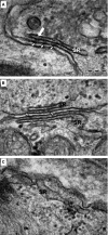
Electron microscopy of peripheral sarcoplasmic reticulum (pSR) in 60–62 days old control, clones KTN3 and HDF24.2 (A), CPVT1, clone 15.4 (B) and CPVT2, clones 20.1 and 19S1 (C) iPSC-CM. Ryanodine receptors (short arrows) are visible between pSR and sarcolemma in control (A) and CPVT1 (B) cardiomyocytes. Enlarged cisterna of pSR is seen in CPVT2 cardiomyocyte (B). Electron-dense linear structure formed by CASQ2 (thick white arrow) is visible within the SR lumen of control cardiomyocytes (A).
Figure 6
Figure 6
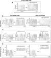
The effects of isoproterenol on the [Ca+2]i transients in control, CPVT1and CPVT2 iPSC-CM. (A) [Ca+2]i transients from control (clone 24.2 day 54) iPSC-CM paced at 0.5 Hz, in the absence and presence of isoproterenol. (B–D) Representative experiments in CPVT1 and CPVT2 iPSC-CM demonstrating the 3 types of responses to isoproterenol. Left side: CPVT1 (clones 15.4 day 56, 15.1 day 49, 15.1 day 38) iPSC-CM; Right side: CPVT2 iPSC-CM (clones 20.1 day 67, 19S1 day 43, 12.1 day 39). (B) No response to isoproterenol. (C) Isoproterenol induced triggered beats, marked by the arrows. (D) Isoproterenol increased diastolic [Ca+2]i. In (B–D) the pacing rates are shown in each panel. Arrhythmias are marked by black arrows.
Figure 7
Figure 7
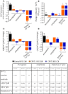
Summary of the effects of isoproterenol on the [Ca2+]i transients of control, CPVT1 and CPVT2 iPSC-CM. (A) [Ca+2]i transient amplitude; (B) Diastolic [Ca2+]i; (C) +d[Ca2+]i/dt; (D) −d[Ca2+]i/dt. In (A, C and D), the effect of isoproterenol was expressed as per cent change from control, and in (B), the effect was expressed as the change in the fluorescence ratio, Δ340/380, *P < 0.05, **P < 0.001. Asterisks above columns represent statistically significant effect of isoproterenol. The statistical analysis of differences among groups is shown in the Table below: C - Control iPSC-CM (n = 8), C1 - CPVT1 iPSC-CM (n = 12), C2 - CPVT2 iPSC-CM (n = 19).
Figure 8
Figure 8
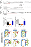
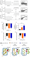
The effect of caffeine on the intracellular Ca2+ cycling of control, CPVT1 and CPVT2 iPSC-CM. (A–C) [Ca2+]i transients from control (clone KTN3 day 68), CPVT1 (clone 15.1 day 53) and CPVT2 (clone 20S3 day 60) iPSC-CM, respectively, demonstrating the effect of caffeine. (D) The per cent change in caffeine-induced Ca2+ signal amplitude, compared to the pre-caffeine [Ca2+]i transient amplitude. (E) The per cent change in area of the caffeine-induced Ca2+ signal, compared to the pre-caffeine [Ca2+]i transient area. Control iPSC-CM (n = 7), CPVT1 iPSC-CM (n = 8), CPVT2 iPSC-CM (n = 8), *P < 0.05, **P < 0.001. Asterisks above columns represent statistically significant effect of caffeine, asterisk above bars connecting columns represent significant difference between groups. (F) A schematic model describing the effects of caffeine in the three groups. In control caffeine depletes SR free Ca2+ stores. Because in CPVT2 there is more (due to the mutated CASQ2) luminal free Ca2+ than in CPVT1 (due to ‘leaky’ RyR2), the response to caffeine is much more pronounced in CPVT2 than in control and CPVT1 iPSC-CM.
Figure 9
Figure 9
The effects of ryanodine on the [Ca2+]i transients of control, CPVT1 and CPVT2 iPSC-CM. (A–C) [Ca2+]i transients from the three groups demonstrating the effect of ryanodine. (D) [Ca+2]i transient amplitude; (E) Diastolic [Ca2+]i; (F) +d[Ca2+]i/dt; (G) −d[Ca2+]i/dt. Control iPSC-CM (n = 11), CPVT1 iPSC-CM (n = 5), CPVT2 iPSC-CM (n = 8). In (D, F and G), the effect of ryanodine was expressed as per cent change from control. In (E), the effect of ryanodine was expressed as the change in the fluorescence ratio ΔF340/380, *P < 0.05, **P < 0.001. Asterisks above columns represent statistically significant effect of ryanodine; asterisk above bars connecting columns represent significant difference between groups. (H) A schematic model explaining the effects of ryanodine in the three groups. Whereas in control cardiomyocytes ryanodine acts as an antagonist and blocks SR Ca2+ release, in the mutated cardiomyocytes, due to altered sensitivity of the deregulated RyR2, ryanodine at 10 μM acts as an agonist, and releases SR Ca2+ into the cytoplasm.

Similar articles

Cited by

References

    1. Kontula K, Laitinen PJ, Lehtonen A, et al. Catecholaminergic polymorphic ventricular tachycardia: recent mechanistic insights. Cardiovasc Res. 2005;67:379–87. - PubMed
    1. Liu N, Priori SG. Disruption of calcium homeostasis and arrhythmogenesis induced by mutations in the cardiac ryanodine receptor and calsequestrin. Cardiovasc Res. 2008;77:293–301. - PubMed
    1. Priori SG, Chen SR. Inherited dysfunction of sarcoplasmic reticulum Ca2+ handling and arrhythmogenesis. Circ Res. 2012;108:871–83. - PMC - PubMed
    1. Fatima A, Xu G, Shao K, et al. In vitro modeling of ryanodine receptor 2 dysfunction using human induced pluripotent stem cells. Cell Physiol Biochem. 2011;28:579–92. - PMC - PubMed
    1. Itzhaki I, Maizels L, Huber I, et al. Modeling of catecholaminergic polymorphic ventricular tachycardia with patient-specific human-induced pluripotent stem cells. J Am Coll Cardiol. 2012;60:990–1000. - PubMed

MeSH terms