. 2015 Jul 8;10(7):e0131894.
doi: 10.1371/journal.pone.0131894. eCollection 2015.

The Effects of the Recombinant CCR5 T4 Lysozyme Fusion Protein on HIV-1 Infection

Affiliations

The Effects of the Recombinant CCR5 T4 Lysozyme Fusion Protein on HIV-1 Infection

Qingwen Jin et al. PLoS One. .

Abstract

Background: Insertion of T4 lysozyme (T4L) into the GPCR successfully enhanced GPCR protein stability and solubilization. However, the biological functions of the recombinant GPCR protein have not been analyzed.

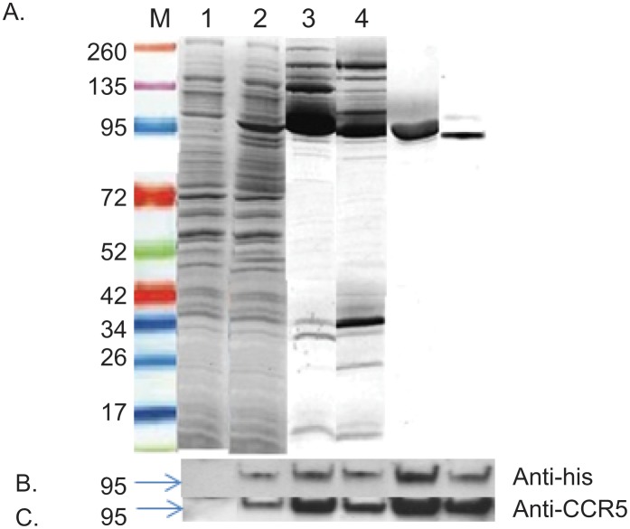
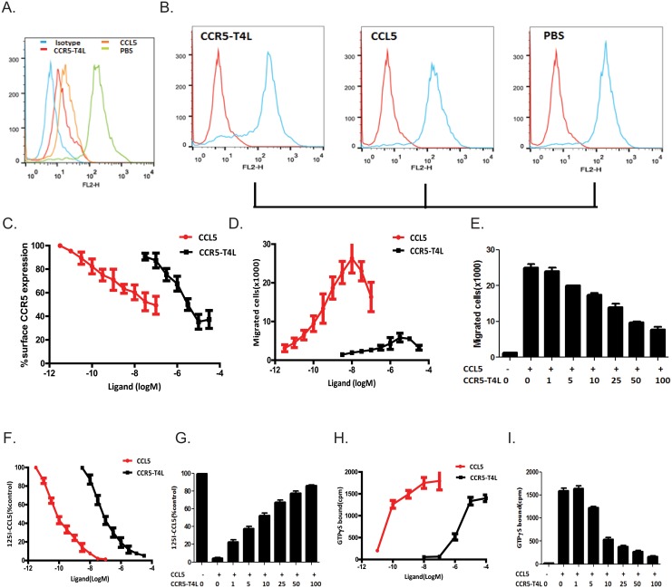
Methods: We engineered the CCR5-T4L mutant and expressed and purified the soluble recombinant protein using an E.coli expression system. The antiviral effects of this recombinant protein in THP-1 cell lines, primary human macrophages, and PBMCs from different donors were investigated. We also explored the possible mechanisms underlying the observed antiviral effects.

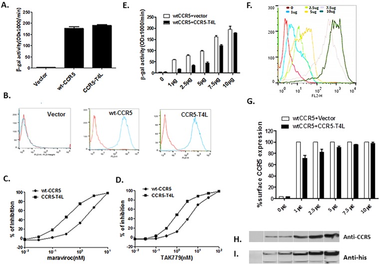
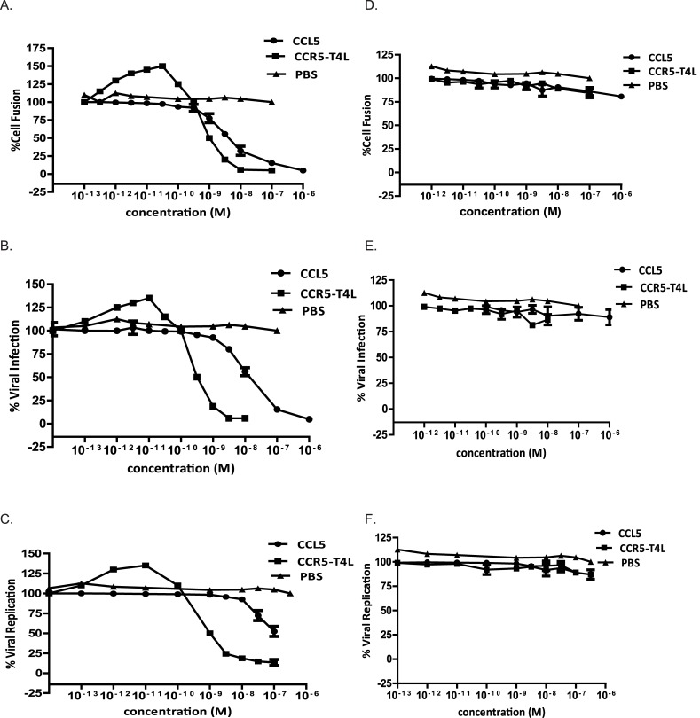
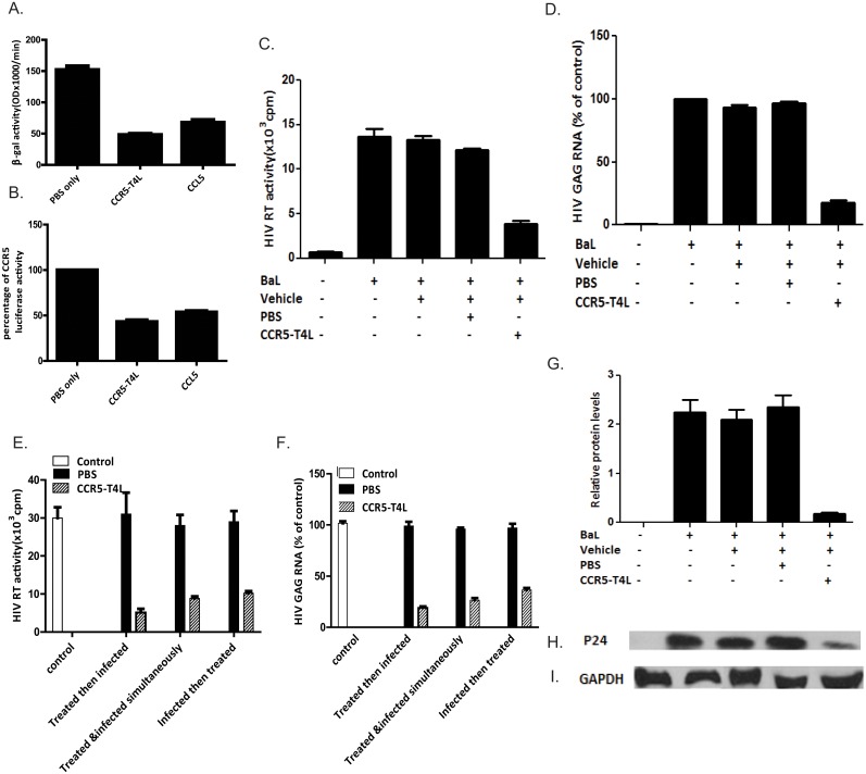
Results: Our data showed the biphasic inhibitory and promotion effects of different concentrations of soluble recombinant CCR5-T4L protein on R5 tropic human immunodeficiency virus-1 (HIV-1) infection in THP-1 cell lines, human macrophages, and PBMCs from clinical isolates. We demonstrated that soluble recombinant CCR5-T4L acts as a HIV-1 co-receptor, interacts with wild type CCR5, down-regulates the surface CCR5 expression in human macrophages, and interacts with CCL5 to inhibit macrophage migration. Using binding assays, we further determined that recombinant CCR5-T4L and [125I]-CCL5 compete for the same binding site on wild type CCR5.

Conclusions: Our results suggest that recombinant CCR5-T4L protein marginally promotes HIV-1 infection at low concentrations and markedly inhibits infection at higher concentrations. This recombinant protein may be helpful in the future development of anti-HIV-1 therapeutic agents.


Conflict of interest statement

Competing Interests: The authors have declared that no competing interests exist.

Figures

Fig 1
Fig 1. Recombinant CCR5-T4L acts as a HIV-1 co-receptor and down-regulates WT-CCR5 expression on the 3T3T4 cell surface.
A) HIV-1 co-receptor function was estimated using cell-cell fusion assays. 3T3.T4 cells were transfected with 15 μg pSC-CCR5-T4L, then infected with vaccinia virus encoding LacZ gene under T7 promotor and used as target cells. The target cells were mixed with HeLa effector cells, expressing BaL Env protein and bacteriophage T7 RNA polymerase. After incubation for 2.5 h at 37°C, the amount of β-galactosidase activity was measured. WT-CCR5 was used as a positive control. B) CCR5 expression on the cell surface was confirmed using FACS. The level of CCR5 expression on above target 3T3.T4 cells was measured by flow cytometry using either phycoerythrin (PE)-conjugated 2D7 or phycoerythrin (PE)-conjugated 3A9 monoclonal antibodies. C and D) CCR5-T4L was more sensitive to CCR5 agonists maraviroc and TAK-779 inhibition. Different concentrations of maraviroc or TAK-779 were used to pretreat target 3T3.T4 cells transfected with pSC-CCR5-T4L or pSC-CCR5 for 1 h at 37°C. After washing with PBS, cell-cell fusion was performed. E and F) CCR5-T4L inhibited WT-CCR5 by reducing its expression on the cell surface. Different amounts of CCR5-T4L or WT-CCR5 cDNA (0 μg、 1 μg、 2.5 μg、5 μg、 7.5 μg and 10 μg) were co-transfected into 3T3.T4 cells. The effect of R5 HIV-1 Env-mediated cell-cell fusion was examined (E) and the level of CCR5 expression on the cell surface was measured by flow cytometry using a PE-conjugated monoclonal antibody (2D7) (F). G) The inhibition by CCR5-T4L decreased as the amounts of CCR5-T4L and WT-CCR5 increased. CCR5 expression was measured using flow cytometry and a PE-conjugated monoclonal antibody (2D7). The averaged mean fluorescence values (MFVs) for CCR5 from three experiments are plotted as a bar diagram, where WT-CCR5 expression after transfection with WT-CCR5 plus empty vector is set at 100. H and I) The interaction between CCR5-T4L and WT-CCR5 was tested using co-immunoprecipitation. 3T3.T4 cells co-transfected with CCR5-T4L and WT-CCR5 were lysed with RIPA buffer. Lysates were prepared and immunoprecipitated with the CCR5 C-terminal antibody, fractionated by SDS-PAGE, and immunoblotted. Blots were probed with the N-terminal CCR5 antibody (H), stripped, and reprobed with the anti-6×His antibody (I). Following the primary antibody reaction, blots were washed and probed with the anti-mouse antibody conjugated to HRP. Blots were exposed to X-ray film after reaction with the HRP substrate.
Fig 2
Fig 2. Expression and purification of soluble recombinant CCR5-T4L protein in an E. coli system.
A) Large scale purification and identification of recombinant CCR5-T4L. Recombinant CCR5-T4L was expressed in E. coli. The pET-20b expression vector was transformed into Rosetta 2 (DE3) golden BL21 pLysS cells and analyzed using Coomassie brilliant blue R-250. Lane M: protein marker; lane 1: uninduced bacterial lysate; lane 2: IPTG-induced bacteria lysate; lane 3: small amount of soluble fraction purified on a Ni-nitrilotriacetic acid (NTA) histidine-binding column; lane 4: small amount of membrane fraction purified using a Ni-nitrilotriacetic acid (NTA) histidine-binding column; lane 5: large amount of soluble fraction purified by fast protein liquid chromatography (FPLC) using an AKTA purifier; lane 6: large amount of membrane fraction purified by fast protein liquid chromatography (FPLC) using an AKTA purifier. B and C) Western blot analyses using the anti-6×His tag monoclonal or anti-human CCR5 monoclonal antibodies (3A9).
Fig 3
Fig 3. Antiviral activities of soluble recombinant CCR5-T4L against R5-tropic and X4-tropic virus.
A and D) The inhibitory activities using cell-cell fusion assays. Effector HeLa cells co-expressing the R5 viral envelope BaL (A) or X4 (D) envelope LAV were pre-incubated for 30 min at 37°C with increasing concentrations of soluble recombinant CCR5-T4L plus 2 nM of soluble CD4. After washing with PBS, cell-cell fusion was performed. B and E) The inhibitory activities using single cycle viral infection assays in THP1 cells. Cells were pre-cultured overnight and infected with BaL or IIIB at 100 TCID50 in the presence or absence of soluble recombinant CCR5-T4L. Luciferase activity was analyzed using a luciferase kit 8 days post-infection. C and F) The inhibitory activities using replication competent viral assays with BaL strain virus- (C) and IIIB strain virus- (F) in THP1 cells. CCR5-tropic HIV-1 ADA was used as a positive control. THP1 cells (1×10⁶ cells per well) were pre-cultured overnight and infected with BaL or IIIB at 100 TCID50 in the presence or absence of soluble recombinant CCR5-T4L overnight. Supernatants were collected 7 days post-infection and tested for p24 antigen using ELISA. Data are the average from three independent experiments. Recombinant ligand CCL5 and PBS were used as controls.
Fig 4
Fig 4. Soluble recombinant CCR5-T4L suppresses HIV infection in macrophages.
A and B) Effect of soluble recombinant CCR5-T4L or CCL5 on macrophages using cell-cell fusion assays (A) and single round virus infection assays (B). Macrophages cultured for 7 days were stimulated for 24 h at 37°C with CCR5-T4L (1 μg/ml) or CCL5 (1 μg/ml) prior to HIV-1 Env-mediated cell-cell fusion or single round virus infection. C, D, G, H, and I) Effect of soluble recombinant CCR5-T4L on HIV BaL infection in macrophages. Macrophages cultured for 7 days were stimulated with CCR5-T4L (1 μg/ml) for 24 h at 37°C prior to HIV-1 Bal infection. Cultured supernatant was collected 8 days post-infection, and cells were collected 12 days post-infection. Supernatants were subjected to RT assays (C), total RNA was evaluated for HIV-1 gag expression using real-time PCR (D), and total protein extracted from cells was evaluated for HIV-1 p24 protein expression by western blot analyses (G and H) and GAPDH (I). Representative blots from three independent experiments are shown. Densitometry analyses of the blot were performed using Image J 1.44 software (NIH) and plotted into graphs (n = 3). E and F) CCR5-T4L suppresses HIV-1 replication in macrophages. Macrophages were cultured at 37°C for 24 h in conditioned media in the presence or absence of CCR5-T4L (1 μg/ml) prior to HIV-1 infection or simultaneously or 8 h post-infection. Supernatants were collected 8 days post-infection, and cells were collected 12 days post-infection. Supernatants were subjected to RT assays (E) and total RNA was evaluated for HIV-1 gag expression using real-time PCR (F). Data are expressed as RNA levels relative to control. The results represent the mean ± SD of three independent experiments.
Fig 5
Fig 5. Recombinant CCR5-T4L down-regulates surface CCR5 expression in MDMs, and inhibits MDM migration and binding properties.
A and B) Effect of soluble recombinant CCR5-T4L or CCL5 on surface CCR5 expression in MDMs. MDMs were treated with soluble recombinant CCR5-T4L (1 μg/ml), CCL5 (1 μg/ml), or PBS for 24 h at 37°C. Surface (A) and intracellular CCR5 (B) were analyzed using flow cytometry and the PE-conjugated monoclonal antibody (2D7). Recombinant CCL5 protein was used as a control. The cellular distribution of CCR5 receptors was analyzed by fixing and permeabilizing cells using BD Cytofix/Cytoperm buffer. Data shown are from one representative experiment that was independently repeated at least three times. C) Dose-dependent effects of CCR5-T4L or CCL5 on surface CCR5 expression in MDM. D and E) Dose-dependent effects on MDM migration by CCR5-T4L (D) or CCL5 (100 nM) plus different concentrations of CCR5-T4L protein (E). F and G) Dose-dependent effects on [125I]-CCR5 binding by CCR5-T4L (F) or CCL5 (100 nM) plus different concentrations of CCR5-T4L protein (G). H and I) Dose-dependent effects on CCL5-induced [35S]GTPγS binding to membranes from human macrophages cells treated with CCR5-T4L (H) or CCL5 (100 nM) plus different concentrations of CCR5-T4L protein (I). Data are the mean ± SD of triplicate cultures, which are representative of three experiments.

Similar articles

Cited by

References

    1. Jones KL, Maguire JJ, Davenport AP. (2011) Chemokine receptor CCR5: from AIDS to atherosclerosis. Br J Pharmacol 162(7):1453–69. 10.1111/j.1476-5381.2010.01147.x - DOI - PMC - PubMed
    1. Klasse PJ (2012) The molecular basis of HIV entry. Cell Microbiol 14(8):1183–92. 10.1111/j.1462-5822.2012.01812.x - DOI - PMC - PubMed
    1. Møller M, Søndergaard HB, Koch-Henriksen N, Sorensen PS, Sellebjerg F, Oturai AB. (2014) The chemokine receptor CCR5 Δ32 allele in natalizumab-treated multiple sclerosis. Acta Neurol Scand 129(1):27–31. 10.1111/ane.12145 - DOI - PubMed
    1. Isgrò M, Bianchetti L, Marini MA, Bellini A, Schmidt M, Mattoli S. (2013) The C-C motif chemokine ligands CCL5, CCL11, and CCL24 induce the migration of circulating fibrocytes from patients with severe asthma. Mucosal Immunol 6(4):718–27. 10.1038/mi.2012.109 - DOI - PubMed
    1. Aiamkitsumrit B, Dampier W, Antell G, Rivera N, Martin-Garcia J, Pirrone V, et al. (2014) Bioinformatic Analysis of HIV-1 Entry and Pathogenesis. Curr HIV Res. 12(2):132–61. - PMC - PubMed

Publication types

MeSH terms