. 2015 Nov;21(11):1188-96.
doi: 10.1089/ten.TEC.2015.0179. Epub 2015 Aug 7.

Embedded 3D Photopatterning of Hydrogels with Diverse and Complex Architectures for Tissue Engineering and Disease Models

Affiliations

Embedded 3D Photopatterning of Hydrogels with Diverse and Complex Architectures for Tissue Engineering and Disease Models

Shruti Krishna Davey et al. Tissue Eng Part C Methods. 2015 Nov.

Abstract

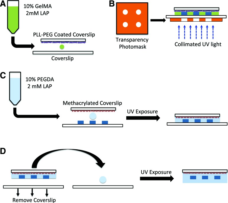
Techniques that can create three-dimensional (3D) structures to provide architectural support for cells have a significant impact in generating complex and hierarchically organized tissues/organs. In recent times, a number of technologies, including photopatterning, have been developed to create such intricate 3D structures. In this study, we describe an easy-to-implement photopatterning approach, involving a conventional fluorescent microscope and a simple photomask, to encapsulate cells within spatially defined 3D structures. We have demonstrated the ease and the versatility of this approach by creating simple to complex as well as multilayered structures. We have extended this photopatterning approach to incorporate and spatially organize multiple cell types, thereby establishing coculture systems. Such cost-effective and easy-to-use approaches can greatly advance tissue engineering strategies.


Figures

<b>FIG. 1.</b>
FIG. 1.
Schematic representation of the protocol for generating three-dimensional (3D) patterned structures. (A) The gelatin methacrylate (GelMA) solution was sandwiched between a poly-L-lysine poly(ethylene glycol)-coated coverslip and a nontreated coverslip. (B) The sandwich was exposed to collimated UV light with a transparency photomask to selectively block the light reaching the GelMA solution. (C) Precursor solution, consisting of poly(ethylene glycol)-diacrylate (PEGDA), was added onto the patterned structures and sandwiched with a methacrylated coverslip before exposing to UV. (D) To completely embed the patterned GelMA structures, PEGDA solution was sandwiched between a coverslip and the previous structure from (C) and exposed to UV. Color images available online at www.liebertpub.com/tec
<b>FIG. 2.</b>
FIG. 2.
Optimization and characterization of patterned structures. (A) The height of the patterned features was adjusted by changing the volume of the GelMA solution from 8 to 20 μL before gelation. Horizontal scale bar: 100 μm. Vertical scale bar: 50 μm. (B) Cylindrical patterns with increasing diameters of 80, 160, and 250 μm were generated by altering the design on the photomask. Scale bar: 75 μm. (C) 3D reconstruction of the GelMA structures with different extruded shapes—circular, triangle, and square. (D) Free-standing patterned GelMA structures surrounded with a PEGDA hydrogel with varying PEGDA concentration (30–10%). The inset shows the GelMA patterns within the PEGDA hydrogel, with the arrow provided for easy identification. Color images available online at www.liebertpub.com/tec
<b>FIG. 3.</b>
FIG. 3.
Generation of complex patterns. GelMA patterns of (A) Sir Isaac Newton and (B) kidney vasculature. Scale bar: 500 μm (C) Confocal sections of bilayer hydrogels containing line patterns showing the X-Z plane and X-Y planes at indicated z positions. Horizontal scale bar: 150 μm. Vertical scale bar: 25 μm. (D) 3D reconstruction of the GelMA structures in the bilayer constructs. Color images available online at www.liebertpub.com/tec
<b>FIG. 4.</b>
FIG. 4.
Encapsulation of cells. (A, B) 3D reconstruction of cells within the patterned hydrogel and the surrounding GelMA hydrogel layer (Left column), where the cells were fluorescently labeled with green cell tracker. (Right column) Phase-contrast and live/dead images of the cylindrical patterns, and live/dead images of distinct layers of the bilayer structures. In live/dead images the green cells indicate viable cells, while the red indicates nonviable cells. Scale bar for (A): 200 μm. Scale bar for (B): 150 μm. (C) Cocultures of human umbilical vein endothelial cell (HUVEC) and MDA-MB-231 cells, where the MDA-MB-231 cell-laden GelMA cylindrical structures are surrounded by hydrogels containing HUVECs. Phase-contrast image (top) along with corresponding live/dead image (bottom). Scale bar: 200 μm. (D) Phase contrast and the fluorescently labeled fibroblasts and HUVECs (bottom) spatially patterned within cylindrical GelMA structures. For easy visualization, the cells were labeled with dyes, with HUVECs represented as red and fibroblasts represented as green. Scale bar: 200 μm. Color images available online at www.liebertpub.com/tec

Similar articles

Cited by

References

    1. Discher D.E., Mooney D.J., and Zandstra P.W. Growth factors, matrices, and forces combine and control stem cells. Science 324, 1673, 2009 - PMC - PubMed
    1. Khetan S., Guvendiren M., Legant W.R., Cohen D.M., Chen C.S., and Burdick J.A. Degradation-mediated cellular traction directs stem cell fate in covalently crosslinked three-dimensional hydrogels. Nat Mater 12, 458, 2013 - PMC - PubMed
    1. Kang H., Shih Y.R., Hwang Y., Wen C., Rao V., Seo T., and Varghese S. Mineralized gelatin methacrylate-based matrices induce osteogenic differentiation of human induced pluripotent stem cells. Acta Biomater 10, 4961, 2014 - PMC - PubMed
    1. Benoit D.S., Schwartz M.P., Durney A.R., and Anseth K.S. Small functional groups for controlled differentiation of hydrogel-encapsulated human mesenchymal stem cells. Nat Mater 7, 816, 2008 - PMC - PubMed
    1. Huebsch N., Arany P.R., Mao A.S., Shvartsman D., Ali O.A., Bencherif S.A., Rivera-Feliciano J., and Mooney D.J. Harnessing traction-mediated manipulation of the cell/matrix interface to control stem-cell fate. Nat Mater 9, 518, 2010 - PMC - PubMed

Publication types