. 2015 Jul 6:4:27378.
doi: 10.3402/jev.v4.27378. eCollection 2015.

Size-exclusion chromatography as a stand-alone methodology identifies novel markers in mass spectrometry analyses of plasma-derived vesicles from healthy individuals

Affiliations

Size-exclusion chromatography as a stand-alone methodology identifies novel markers in mass spectrometry analyses of plasma-derived vesicles from healthy individuals

Armando de Menezes-Neto et al. J Extracell Vesicles. .

Abstract

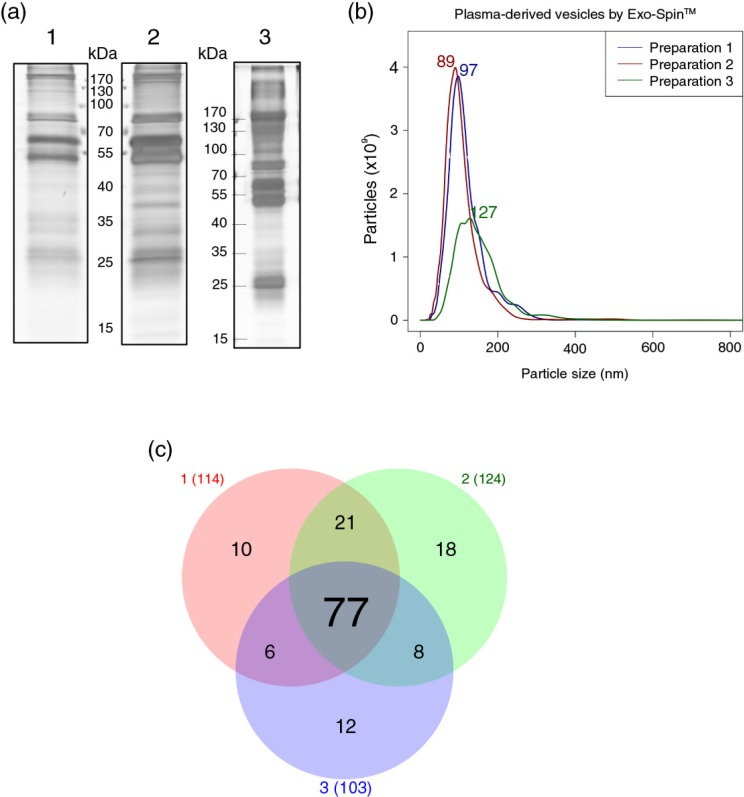
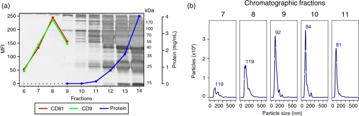
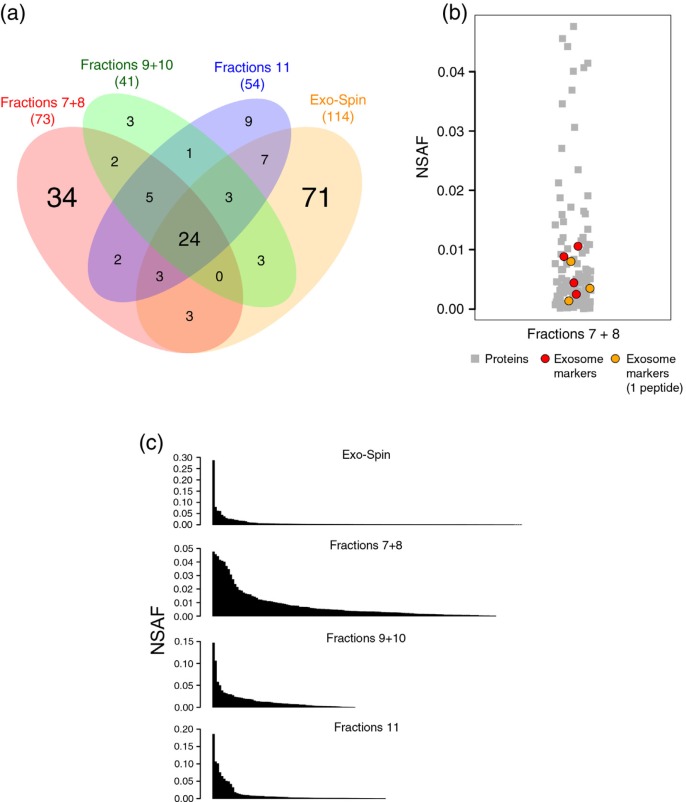
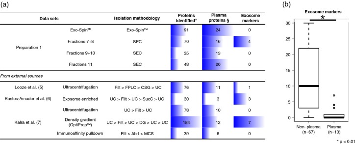
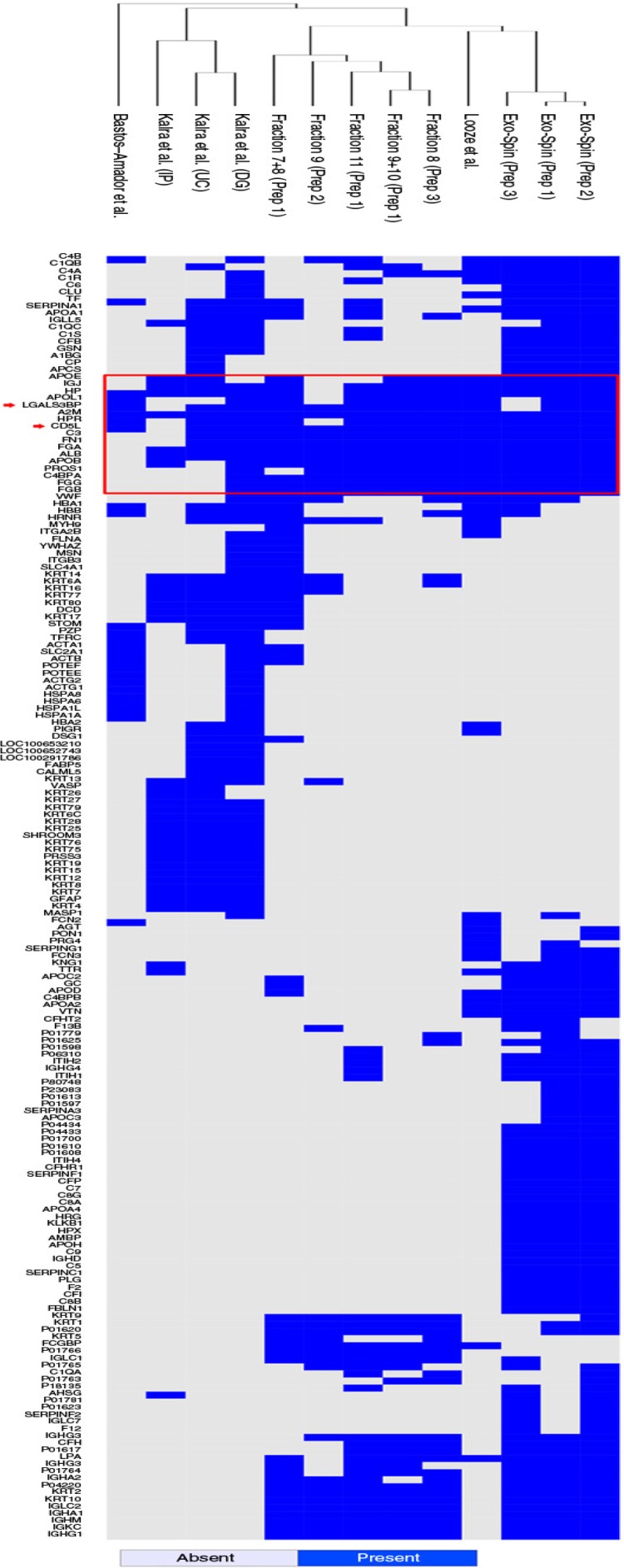
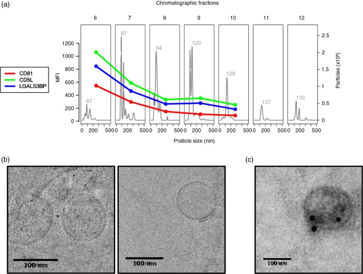
Plasma-derived vesicles hold a promising potential for use in biomedical applications. Two major challenges, however, hinder their implementation into translational tools: (a) the incomplete characterization of the protein composition of plasma-derived vesicles, in the size range of exosomes, as mass spectrometric analysis of plasma sub-components is recognizably troublesome and (b) the limited reach of vesicle-based studies in settings where the infrastructural demand of ultracentrifugation, the most widely used isolation/purification methodology, is not available. In this study, we have addressed both challenges by carrying-out mass spectrometry (MS) analyses of plasma-derived vesicles, in the size range of exosomes, from healthy donors obtained by 2 alternative methodologies: size-exclusion chromatography (SEC) on sepharose columns and Exo-Spin™. No exosome markers, as opposed to the most abundant plasma proteins, were detected by Exo-Spin™. In contrast, exosomal markers were present in the early fractions of SEC where the most abundant plasma proteins have been largely excluded. Noticeably, after a cross-comparative analysis of all published studies using MS to characterize plasma-derived exosomes from healthy individuals, we also observed a paucity of "classical exosome markers." Independent of the isolation method, however, we consistently identified 2 proteins, CD5 antigen-like (CD5L) and galectin-3-binding protein (LGALS3BP), whose presence was validated by a bead-exosome FACS assay. Altogether, our results support the use of SEC as a stand-alone methodology to obtain preparations of extracellular vesicles, in the size range of exosomes, from plasma and suggest the use of CD5L and LGALS3BP as more suitable markers of plasma-derived vesicles in MS.

Keywords: CD5L; LGALS3BP; comparative analysis; exosomes; low infrastructure settings; mass spectrometry; plasma-derived exosomes.


Figures

Fig. 1
Fig. 1
Plasma-derived exosomes isolated by Exo-Spin™ from the plasma of 3 healthy donors. (a) Electrophoretic profile of proteins on silver-stained polyacrylamide gels. The following amount of proteins were loaded onto the gels: 1.05 µg (1); 1.14 µg (2) and 4.6 µg (3). (b) NTA was performed on a NanoSight LM10 (software version 2.3) after dilution of samples in PBS. (c) Venn diagram showing the overlap of proteins detected by LC–MS/MS in each preparation (red: “Preparation from donor 1”; green: “Preparation from donor 2”; blue: “Preparation from donor 3”).
Fig. 2
Fig. 2
Isolation of plasma-derived exosomes by size-exclusion chromatography. An aliquot (1 mL) of undiluted plasma from Donor 1 was passed through a sepharose (CL-2B) column, and 30 fractions of 0.5 mL each were collected. (a) SDS–PAGE stained with silver and protein concentration values of fractions 6–14 were measured by Bradford assay (fractions 6, 7 and 8 were below the lower limit of detection) and fractions 6–9 were analysed by flow cytometry, after coupling of vesicles to 4 µm latex beads, for the presence of antigens CD9 (1:10) and CD81 (1:10). The secondary anti-mouse antibody was conjugated to FITC and was used at a 1:100 dilution. MFI: mean fluorescence intensity. (b) Fractions 7–11 were submitted to NTA on a NanoSight LM10 (software version 3.0).
Fig. 3
Fig. 3
Proteomic analysis of samples from donor 1. Mass spectrometry was performed on 4 preparations from the same plasma sample: (i) chromatographic fractions 7+8, (ii) chromatographic fractions 9+10, (iii) chromatographic fraction 11 and (iv) Exo-Spin™. (a) Venn diagram showing the overlap of proteins detected by nanoLC–MS/MS. (b) The relative abundance (NSAF) of the exosome markers detected in the data set of fractions 7+8 (in red) and of exosome markers identified by single peptides (orange) were compared to the relative abundance of the remaining proteins for the same sample. (c) The overall relative abundance of all proteins in each sample plotted in descending order. NSAF=normalized spectrum abundance factor.
Fig. 4
Fig. 4
Exosome markers in plasma-derived exosomal preparations. (a) Mass spectrometry results from Exo-Spin and SEC samples (Preparation 1) were compared to previous studies that have isolated exosomes by other methodologies. The total number of identified proteins, of abundant plasma proteins and of exosome markers, is shown. (b) A panel of 34 exosome markers are compared between high-throughput proteomic data sets of plasma-derived exosomes (n = 13) and non-plasma-derived exosomes retrieved from ExoCarta (n = 67). Statistical significance was tested for with Mann–Whitney–Wicoxon test for non-parametric distributions at a level of significance of 5% (p-value < 0.05). SEC: size-exclusion chromatography, FPLC: fast protein liquid chromatography, SCG: continuous sucrose gradient, UC: ultracentrifugation, Filt: filtration at 0.22 or 0.1 µm, SucC: sucrose cushion, DG: density gradient, Ab-I: antibody incubation, MCS: magnetic column separation, ¥according to data extracted from ExoCarta for previous publications, §list of common plasma proteins.
Fig. 5
Fig. 5
Hierarchical clustering of plasma data sets. The 13 data sets of proteomic data on plasma-derived exosomes were clustered after removal of proteins that were detected in only one of the data sets. For the remaining 182 proteins, a distance matrix based on their presence/absence was built using a binary method. The calculated distances were used to generate the hierarchical cluster. The red rectangle highlights the proteins that were more frequently detected (≥9). The red arrows indicate 2 proteins, CD5 antigen-like (CD5L) and galectin-3 binding protein (LGALS3BP).
Fig. 6
Fig. 6
CD5L and LGALS3BP have similar elution profiles to the exosome marker CD81. Plasma from “Donor 1” was submitted to size-exclusion chromatography and fractions 6–12 were analysed by NTA, flow cytometry and transmission electron microscopy (TEM). (a) NTA was performed on a NanoSight LM10 (software version 3.0). For flow cytometry, samples were coupled to 4 µm beads and incubated with primary antibodies against CD81 (1:10), CD5L (1:100) or LGALS3BP (1:1,000). The secondary antibody was conjugated to Alexa 488 was used at a 1:1,000 dilution. MFI: mean fluorescence intensity. (b) Fraction 6 from size-exclusion chromatography was submitted to cryo-EM and (c) immunostained with anti-CD5L antibodies conjugated to gold spheres of 20 nm.

References

    1. Anderson NL. The human plasma proteome: history, character, and diagnostic prospects. Mol Cell Proteomics. 2002;1:845–67. doi: 10.1074/mcp.R200007-MCP200. - DOI - PubMed
    1. Revenfeld ALS, Bæk R, Nielsen MH, Stensballe A, Varming K, Jørgensen M. Diagnostic and prognostic potential of extracellular vesicles in peripheral blood. Clin Ther. 2014;36:830–46. doi: 10.1016/j.clinthera.2014.05.008. - DOI - PubMed
    1. Théry C, Zitvogel L, Amigorena S. Exosomes: composition, biogenesis and function. Nat Rev Immunol. 2002;2:569–79. doi: 10.1038/nri855. - DOI - PubMed
    1. Valadi H, Ekström K, Bossios A, Sjöstrand M, Lee JJ, Lötvall JO. Exosome-mediated transfer of mRNAs and microRNAs is a novel mechanism of genetic exchange between cells. Nat Cell Biol. 2007;9:654–9. doi: 10.1038/ncb1596. - DOI - PubMed
    1. Looze C, Yui D, Leung L, Ingham M, Kaler M, Yao X, et al. Proteomic profiling of human plasma exosomes identifies PPARgamma as an exosome-associated protein. Biochem Biophys Res Commun. 2009;378:433–8. doi: 10.1016/j.bbrc.2008.11.050. - DOI - PMC - PubMed