. 2015 Jul 8:6:7646.
doi: 10.1038/ncomms8646.

Cryo-EM structure of Hepatitis C virus IRES bound to the human ribosome at 3.9-Å resolution

Affiliations

Cryo-EM structure of Hepatitis C virus IRES bound to the human ribosome at 3.9-Å resolution

Nick Quade et al. Nat Commun. .

Abstract

Hepatitis C virus (HCV), a widespread human pathogen, is dependent on a highly structured 5'-untranslated region of its mRNA, referred to as internal ribosome entry site (IRES), for the translation of all of its proteins. The HCV IRES initiates translation by directly binding to the small ribosomal subunit (40S), circumventing the need for many eukaryotic translation initiation factors required for mRNA scanning. Here we present the cryo-EM structure of the human 40S ribosomal subunit in complex with the HCV IRES at 3.9 Å resolution, determined by focused refinement of an 80S ribosome-HCV IRES complex. The structure reveals the molecular details of the interactions between the IRES and the 40S, showing that expansion segment 7 (ES7) of the 18S rRNA acts as a central anchor point for the HCV IRES. The structural data rationalizes previous biochemical and genetic evidence regarding the initiation mechanism of the HCV and other related IRESs.


Figures
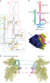
Figure 1
Figure 1. HCV IRES bound to human ribosome.
(a) Secondary structure diagram of the HCV IRES. The various domains are indicated by different colours and roman numbers. Canonical base pairs are marked by lines and non-standard base pairs by circles. Interactions of the IRES with ribosomal proteins are highlighted in orange, interactions with the 18S rRNA in dark yellow. Bases not built in our structure are indicated in grey, the start codon is indicated by a black box. (b) Cartoon representation of the HCV IRES secondary structure. The interaction sites of the different domains are indicated. (c) EM density of 80S–HCV IRES complex showing that the HCV IRES does not contact the 60S subunit. The 40S is coloured in yellow, the 60S in blue and the HCV IRES in pink. (d) Structure of the HCV IRES bound to the human 40S. Views from the intersubunit side (left) and the solvent side (right).
Figure 2
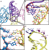
Figure 2. ES7 acts as the main anchoring point for the HCV IRES.
(a) Overview over the architecture of the binding site. (b,c) Close-ups of the interactions between ES7 and domain IIId (b) and IIIe (c). The structures are shown in cartoon and sticks, red dashes indicate hydrogen bonds. (d) Conformation of ES7 bound to HCV IRES. Bases interacting with domain IIId are coloured in purple, and bases interacting with domain IIIef are coloured in dark blue.
Figure 3
Figure 3. Domain IIIac binding site on eS27.
Orange spheres indicate amino acids interacting with the IRES. Inset panel: detailed interactions between bases G163 and G233 of domain IIIac with amino acids from eS27. The structure is shown as cartoon, interacting residues as sticks.
Figure 4
Figure 4. The HCV IRES and eIF3 occupy distinct binding sites on the 40S.
Ribosomal proteins are coloured in yellow, the bases of the 18S rRNA are coloured in light grey, the backbone in dark grey. eIF3-binding sites are shown in orange, HCV IRES binding sites are coloured according to their interacting domain (green: domain II, blue: domain IIIef, purple: domain IIId and pink: domain IIIac). The figure was generated by fitting the structure of the 40S bound to the HCV IRES into the density of 40S bound to eIF3 (ref. 26) (EMDB ID 5658).
Figure 5
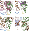
Figure 5. Interactions between ribosomal proteins and domain IIIf and domain II of the HCV IRES.
(a) Overview over HCV binding sites. Protein uS7 is shown in ochre, uS11 in pink, eS25 in light blue and eS28 in light green. Other ribosomal proteins are coloured in yellow, 18S rRNA in grey and domain II and IIef in green and blue, respectively. Interacting residues are marked by spheres. (bd) Detailed views of binding sites of domain IIIf to eS28 (b), the apical domain of domain II to uS7 and uS11 (c) and a minor interaction site between domain II and uS7 and eS25 (d). Structures are depicted as cartoon. The dashed blue line indicates the approximate path of the mRNA.
Figure 6
Figure 6. HCV IRES domain II occupies similar binding sites as E-site tRNA, eIF2 and CrPV IRES.
(a) The apical loop of domain II binds to the β-hairpin of uS7 similar to E-site tRNA. Superposition of the HCV IRES bound to 40S with a bacterial ribosome containing an E-site tRNA (PDB ID 4RBH) based on uS7. The structure of the bacterial ribosome is shown in red, in the HCV structure domain II is coloured green, mRNA in blue and uS7 in ochre. (b) Superposition of the HCV IRES-80S structure and a partial 48S preinitiation complex (PDB ID 3J81) on uS7 illustrating that eIF2 binding and binding of domain II at the E-site are mutually exclusive. (c) Comparison of uS7 interactions of the HCV IRES bound to 80S with the CrPV IRES bound to 80S (coloured in brown). (d) Comparison of eS25 interactions of the HCV IRES bound to 80S with CrPV IRES bound to 80S (coloured in brown). The dashed blue line indicates the approximate path of the mRNA.

References

    1. Gravitz L. Introduction: a smouldering public-health crisis. Nature 474, S2–S4 (2011). - PubMed
    1. Hinnebusch A. G. The scanning mechanism of eukaryotic translation initiation. Annu. Rev. Biochem. 83, 779–812 (2014). - PubMed
    1. Kieft J. S., Zhou K., Jubin R. & Doudna J. A. Mechanism of ribosome recruitment by hepatitis C IRES RNA. RNA 7, 194–206 (2001). - PMC - PubMed
    1. Pestova T. V., Shatsky I. N., Fletcher S. P., Jackson R. J. & Hellen C. U. A prokaryotic-like mode of cytoplasmic eukaryotic ribosome binding to the initiation codon during internal translation initiation of hepatitis C and classical swine fever virus RNAs. Genes Dev. 12, 67–83 (1998). - PMC - PubMed
    1. Otto G. A. & Puglisi J. D. The pathway of HCV IRES-mediated translation initiation. Cell 119, 369–380 (2004). - PubMed

Publication types

MeSH terms

Substances

Associated data