

Case report of a pancreatic squamoid cyst
- PMID: 26155237
- PMCID: PMC4304511
- DOI: 10.14701/kjhbps.2013.17.4.181
Case report of a pancreatic squamoid cyst
Abstract
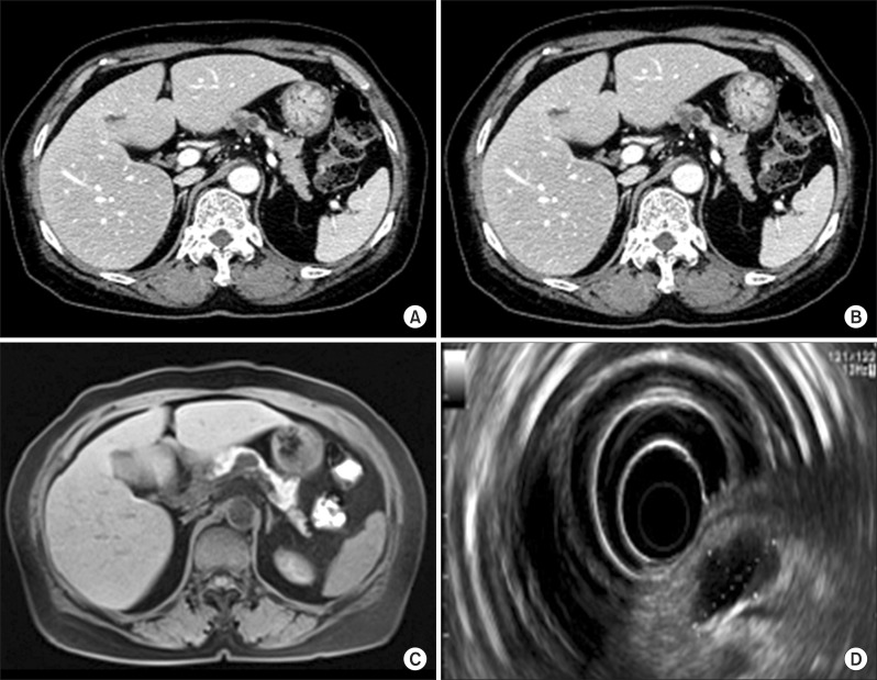
Squamoid cyst of the pancreas is a very rare disease and it has been proposed only recently as a distinct pathologic lesion. We herein present a case of pancreatic squamoid cyst in a patient who underwent laparoscopic resection. A 60-year-old woman had an abdominal computed tomography (CT) scan for a routine check-up, and a multi-cystic lesion of 1.8-cm in size was incidentally found in the tail of the pancreas. Biochemical laboratory tests were within normal limits. At first, we presumed that the most likely diagnosis of the cystic lesion was an intraductal papillary mucinous neoplasm. To treat this lesion, we performed laparoscopic spleen-saving distal pancreatectomy. The patient showed the usual routine postoperative course and she was discharged 10 days after surgery. On examination of the resected specimen, a well-defined, oligolocular cystic mass was found in the pancreatic tail, without a solid portion. Histologic examination revealed that the cysts had linings ranging from flat squamoid cells to transitional cells with non-keratinization. After immunohistochemical staining, the final diagnosis was confirmed to be squamoid cyst of the pancreas. This lesion appears to be regarded as a benign entity, thus an extended operation should be avoided and resection of the lesion can be performed minimally.
Keywords: Pancreatic cancer; Pancreatic squamoid cyst.
Figures


Similar articles
-
Squamoid cyst of pancreatic ducts: A case report.Ann Hepatobiliary Pancreat Surg. 2021 May 31;25(2):293-298. doi: 10.14701/ahbps.2021.25.2.293. Ann Hepatobiliary Pancreat Surg. 2021. PMID: 34053935 Free PMC article.
-
Squamoid cystosis of pancreatic ducts: a variant of a newly-described cystic lesion, with evidence for an obstructive etiology.Rare Tumors. 2014 Sep 17;6(3):5286. doi: 10.4081/rt.2014.5286. eCollection 2014 Jul 30. Rare Tumors. 2014. PMID: 25276318 Free PMC article.
-
[Two cases of squamoid cyst of the pancreatic ducts].Nihon Shokakibyo Gakkai Zasshi. 2015 May;112(5):896-904. doi: 10.11405/nisshoshi.112.896. Nihon Shokakibyo Gakkai Zasshi. 2015. PMID: 25947026 Review. Japanese.
-
Squamoid cyst of pancreatic ducts: a challenging differential diagnosis among benign pancreatic cysts.JOP. 2013 Nov 10;14(6):657-60. doi: 10.6092/1590-8577/1905. JOP. 2013. PMID: 24216555
-
Laparoscopic distal pancreatectomy for a pancreatic lymphoepithelial cyst: case report and review of literature.JOP. 2013 Nov 10;14(6):664-8. doi: 10.6092/1590-8577/1738. JOP. 2013. PMID: 24216557 Review.
Cited by
-
Intraductal papillary squamous neoplasm of the pancreas: Cyto-histologic correlation of a novel entity.Ann Diagn Pathol. 2020 Oct;48:151583. doi: 10.1016/j.anndiagpath.2020.151583. Epub 2020 Aug 13. Ann Diagn Pathol. 2020. PMID: 32847795 Free PMC article.
-
Squamoid cyst of pancreatic ducts: A case report.Ann Hepatobiliary Pancreat Surg. 2021 May 31;25(2):293-298. doi: 10.14701/ahbps.2021.25.2.293. Ann Hepatobiliary Pancreat Surg. 2021. PMID: 34053935 Free PMC article.
References
-
- Kurahara H, Shinchi H, Mataki Y, et al. A case of squamoid cyst of pancreatic ducts. Pancreas. 2009;38:349–351. - PubMed
-
- Othman M, Basturk O, Groisman G, et al. Squamoid cyst of pancreatic ducts: A distinct type of cystic lesion in the pancreas. Am J Surg Pathol. 2007;31:291–297. - PubMed
-
- Brugge WR. The use of EUS to diagnose cystic neoplasms of the pancreas. Gastrointest Endosc. 2009;69(2 Suppl):S203–S209. - PubMed
-
- Adsay NV, Klimstra DS, Compton CC. Cystic lesions of the pancreas. Introduction. Semin Diagn Pathol. 2000;17:1–6. - PubMed
-
- Kanazawa H, Kamiya J, Nagino M, et al. Epidermoid cyst in an intrapancreatic accessory spleen: a case report. J Hepatobiliary Pancreat Surg. 2004;11:61–63. - PubMed
Publication types
LinkOut - more resources
Full Text Sources
Other Literature Sources