Review
. 2014;4(6):169-88.
doi: 10.1080/19490992.2015.1054092.

Myosin light chains: Teaching old dogs new tricks

Affiliations
Review

Myosin light chains: Teaching old dogs new tricks

Sarah M Heissler et al. Bioarchitecture. 2014.

Abstract

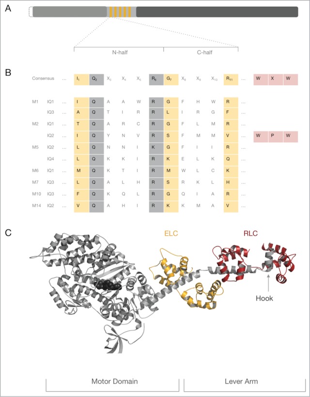
The myosin holoenzyme is a multimeric protein complex consisting of heavy chains and light chains. Myosin light chains are calmodulin family members which are crucially involved in the mechanoenzymatic function of the myosin holoenzyme. This review examines the diversity of light chains within the myosin superfamily, discusses interactions between the light chain and the myosin heavy chain as well as regulatory and structural functions of the light chain as a subunit of the myosin holoenzyme. It covers aspects of the myosin light chain in the localization of the myosin holoenzyme, protein-protein interactions and light chain binding to non-myosin binding partners. Finally, this review challenges the dogma that myosin regulatory and essential light chain exclusively associate with conventional myosin heavy chains while unconventional myosin heavy chains usually associate with calmodulin.

Keywords: actin; calmodulin; cytoskeleton; essential light chain; live cell imaging; myosin; regulatory light chain.


Figures

Figure 1.
Figure 1.
The myosin IQ motif in the heavy chain. (A) Schematic representation of a generic myosin heavy chain. The N-terminus of the heavy chain represents the catalytic motor domain (gray) that harbors the nucleotide binding site and the F-actin binding region. The central neck domain harbors the IQ motifs (orange). The C-terminal tail domain (dark gray) can contain a vast collection of domains and motifs that determine the oligomeric state of the heavy chain and interaction signatures with binding partners. The length of the myosin motor domain is very conserved, whereas the length of the myosin neck and tail domain varies considerably. Some myosins have an N-terminal extension of variable length prior to the motor domain which is indicated by the dashed line. (B) The top line represents the consensus IQ motif found in myosin heavy chains. Position 1 of the 11 amino acid long consensus IQ motif is critically occupied by a hydrophobic residue such as isoleucine (I), leucine (L) or valine (V) though methionine (M), phenylalanine (F) and lysine (K) and threonine (T) are found in some cases. Position 7 is ambiguous for several amino acids. Position 11 is preferentially occupied by a positively charged amino acid (arginine (R), histidine (H) or lysine (K)). Invariant residues are boxed in gray, ambiguous residues in peach. Non-boxed residues are highly variable. The WXW motif found in the hook region of conventional myosins-2 is colored in rose. Of note, the WXW motif is not part of the consensus IQ motif but crucial for the interaction of RLCs with the IQ2 in myosins-2 and therefore depicted for completeness. All other lines shown represent selected IQ motif found in conventional and unconventional myosin heavy chains discussed in this review. The abbreviations used are as follows: M1: IQ1 and IQ3 from human myosin-1C, M2: IQ1 and IQ2 from chicken striated muscle myosin-2; M5: IQ2 and IQ4 from budding yeast Myo2p; M6: IQ1 from pig myosin-6; M7: IQ3 from human myosin-7A; M10: IQ3 from human myosin-10; M14: IQ2 from P.yoelli. (C) Crystal structure of a prototypic myosin-2–2IQ fragment. Structural overview of the striated myosin-2–2IQ fragment from scallop (PDB ID: 1DFL). The myosin heavy chain is shown in gray cartoon representation, the nucleotide in black spheres. The associated ELC and RLC are represented in orange and cherry color, respectively. ELC and RLC bind distal from the globular motor domain to the IQ motifs of the neck domain. The myosin neck region and the associated light chains form the lever arm. The hook is indicated by a gray arrow.
Figure 2.
Figure 2.
Structure of CaM and its EF-hand motifs in various conformations. (A) Structures of CaM (blue) bound to a target sequence (gray) in the extended (PDB ID: 3CLN) and compact conformation (PDB ID: 2IX7). (B) EF-hand conformations found in CaM. Top row: EF-hand in an open (PDB ID: 1CLL) or closed (PDB ID: 1CFC) conformation. Bottom row: The semi-open (PDB ID 2IX7) and the uncoupled conformation of the EF-hand motif (PDB ID 4R8G). Figure 2B is adapted from Lu et al.
Figure 3.
Figure 3.
Structural consequences of RLC phosphorylation on the conformation of smooth and nonmuscle myosins-2. RLC phosphorylation at S19 promotes a conformational change from a compact (left) to an extended conformation of the myosin holoenzyme (left), which readily assembles into higher order bipolar filaments (not shown). The structural transition from the compact to the expended conformation is accompanied with an increase in the mechanoenzymatic activity. RLC dephosphorylation by myosin phosphatase reverses the conformational change. Figure adapted from Heissler and Manstein.
Figure 4.
Figure 4.
Consequences of different binding modes of myosin light chains. (A) Schematic representation of the open and closed conformation of CaM (blue) bound to an exclusive target IQ-motif (gray). CaM-dependent dimerization of 2 domains of the Ca2+-activated K+ channel (gray), each bound to one lobe of CaM (PDB ID: 1G4Y). (B) Extended and compact light chain conformation exemplarily shown for the ELC (orange) on the IQ motifs (gray) of the yeast myosin-5 (PDB IDs: 1M45, 1M46). Potential functional consequences of the extended and compact light chain conformation are valid for ELC, RLC and CaM.

References

    1. Sellers JR. Myosins: a diverse superfamily. Biochim Biophys Acta 2000; 1496:3-22; PMID:10722873; http://dx.doi.org/10.1016/S0167-4889(00)00005-7 - DOI - PubMed
    1. Odronitz F, Kollmar M. Drawing the tree of eukaryotic life based on the analysis of 2,269 manually annotated myosins from 328 species. Genome Biol 2007; 8:R196; PMID:17877792; http://dx.doi.org/10.1186/gb-2007-8-9-r196 - DOI - PMC - PubMed
    1. Cheney RE, Mooseker MS. Unconventional myosins. Curr Opin Cell Biol 1992; 4:27-35; PMID:1558751; http://dx.doi.org/10.1016/0955-0674(92)90055-H - DOI - PubMed
    1. Bahler M, Rhoads A. Calmodulin signaling via the IQ motif. FEBS Lett 2002; 513:107-13; PMID:11911888; http://dx.doi.org/10.1016/S0014-5793(01)03239-2 - DOI - PubMed
    1. Vetter SW, Leclerc E. Novel aspects of calmodulin target recognition and activation. Eur J Biochem 2003; 270:404-14; PMID:12542690; http://dx.doi.org/10.1046/j.1432-1033.2003.03414.x - DOI - PubMed

Publication types

LinkOut - more resources