. 2015 Aug 6;12(109):20150342.
doi: 10.1098/rsif.2015.0342.

Cell-cell signalling in sexual chemotaxis: a basis for gametic differentiation, mating types and sexes

Affiliations

Cell-cell signalling in sexual chemotaxis: a basis for gametic differentiation, mating types and sexes

Zena Hadjivasiliou et al. J R Soc Interface. .

Abstract

While sex requires two parents, there is no obvious need for them to be differentiated into distinct mating types or sexes. Yet this is the predominate state of nature. Here, we argue that mating types could play a decisive role because they prevent the apparent inevitability of self-stimulation during sexual signalling. We rigorously assess this hypothesis by developing a model for signaller-detector dynamics based on chemical diffusion, chemotaxis and cell movement. Our model examines the conditions under which chemotaxis improves partner finding. Varying parameter values within ranges typical of protists and their environments, we show that simultaneous secretion and detection of a single chemoattractant can cause a multifold movement impediment and severely hinder mate finding. Mutually exclusive roles result in faster pair formation, even when cells conferring the same roles cannot pair up. This arrangement also allows the separate mating types to optimize their signalling or detecting roles, which is effectively impossible for cells that are both secretors and detectors. Our findings suggest that asymmetric roles in sexual chemotaxis (and possibly other forms of sexual signalling) are crucial, even without morphological differences, and may underlie the evolution of gametic differentiation among both mating types and sexes.

Keywords: chemotaxis; mating types; sexes; signalling.


Figures

Figure 1.
Figure 1.
Chemical concentration around moving secretors. Cells secrete a diffusible chemical (red and blue diamonds) that binds to membrane receptors thereby inducing a chemotactic signal. Molecules secreted by the cell can cause two problems. First, they bind to the cell's own receptors causing saturation and interference with signals from remote partners whose molecules are always at relatively low concentration because of diffusion (the red cell has most of its receptors occupied by its own pheromone). Second, owing to cell movement, secretion causes a tail of high concentration behind the moving cell. It follows that receptor occupancy is higher behind the moving cells, prompting the cell to repeatedly reverse direction (the blue cell's receptors are occupied mainly at its rear).
Figure 2.
Figure 2.
Chemotactic cells change their direction according to the chemical gradient. The vector g shown in red is a unit vector along the direction of the gradient. The cell updates its position by taking a step of length l along the direction of the dotted green vector which is the sum of a unit vector along a random direction and a magnified vector along the direction of the gradient. The greater this magnification (determined by αΔB), the closer is the direction the cell moves in to the direction of the gradient. l is chosen from a uniform distribution on [0, 2].
Figure 3.
Figure 3.
High persistence minimizes half-life of non-chemotactic and secrete-and-detect cells. (a) Mean half-life (averaged over 40 simulations) against persistence for non-chemotactic cells (blue) and secrete-and-detect cells (black). Example trajectories of two cells until they meet for (b–d) non-chemotactic cells and (e–g) secrete-and-detect cells. Initial positions are spaced equally far apart (blue dots). The persistence parameter for each simulation is indicated at the top of each plot. The duration of the search (t) is given at the bottom of each square. Other parameters: (ρ0, s, s/Kd, u, d, α, v) = (5.1 × 10⁶ cells m−2, 1 s−1, 10−4, 10−3 s−1, 40 µm, 100, 100 µm s−1).
Figure 4.
Figure 4.
Low persistence minimizes half-life for separate secrete-only and detect-only cells. (a) Heat map of the mean half-life (averaged over 40 simulations) for secrete-only and detect-only cells (hS+D) given different values of persistence for secretors (pS) and detectors (pD). (b) The half-life (mean ± s.d. of 40 simulations) for secrete-only and detect-only cells (red) is compared with the half-life of secrete-and-detect cells for variable persistence p (black) for fixed secretor (pS = 0.9) and variable detector persistence, and (c) for fixed detector (pD = 0.9) and variable secretor persistence. (d–f) Example trajectories of two cells, one secretor and one detector, given different pS and pD values. Initial positions are spaced equally far apart (blue dots), with the duration of the search (t) given at the bottom of each square. Other parameters: (ρ0, s, u, d, α, v) = (5.1 × 10⁶ cells m−2, 1 s−1, 10−3 s−1, 40 µm, 100, 100 µm s−1). The ratio s/Kd is set equal to 10−4 and 10−5 for SD and S + D cells, respectively.
Figure 5.
Figure 5.
Diffusion and speed. Mean half-life ratios with error bars (averaged over 40 simulations) for secrete-only and detect-only cells (red) compared with non-chemotactic (blue dotted) and secrete-and-detect (black) cells, for varying cell speed given (a) high diffusion Dc = 10−5 cm2 s−1 and (b) low diffusion Dc = 10−6 cm2 s−1. Values below the blue line indicate that chemotaxis confers an improvement in the rate of pair formation. The asymmetry of chemical concentration around a moving secretor (direction indicated by the arrow) is greater as the secretor's speed (v) increases and the chemical diffusivity (Dc) decreases (illustrated in a single dimension in (c) and (d), respectively). Other parameters (ρ0, s, u, d, α) = (5.1 × 10⁶ cells m−2, 1 s−1, 10−3 s−1, 40 µm, 100). The ratio s/Kd is set equal to 10−4 and 10−5 for SD and S + D cells, respectively. The persistence parameter for NC and SD cells is set equal to 0.2. For S + D cells, we set (pS, pD) = (0.2, 0.5).
Figure 6.
Figure 6.
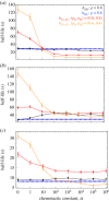
The half-life against the chemotactic constant. The half-life for secrete-only and detect-only cells (red and orange) compared with non-chemotactic (blue dotted), and secrete-and-detect (black) cells (mean ± s.d. of 40 simulations), for variation in the chemotactic constant (α) under (a) intermediate cell speed v = 100 µm s−1 and high diffusion coefficient Dc = 10−5 cm2 s−1, (b) intermediate cell speed v = 100 µm s−1 and low diffusion coefficient Dc = 10−6 cm2 s−1 and (c) high cell speed v = 250 µm s−1 and high diffusion coefficient Dc = 10−5 cm2 s−1. Non-chemotactic cells are not affected by the value of α. Values below the blue line indicate that chemotaxis confers an improvement in the rate of pair formation. Other parameters: (ρ0, s, u, d) = (5.1 × 10⁶ cells m−2, 1 s−1, 10−3 s−1, 40 µm). The ratio s/Kd is set equal to 10−4 and 10−5 for SD and S + D cells, respectively.
Figure 7.
Figure 7.
The effect of cell size. The half-life of non-chemotactic (blue line), secrete-and-detect cells (black line) and for secrete-only and detect-only cells (red line) against cell diameter (mean ± s.d. of 40 simulations). Other parameters: (ρ0, s, u, d, α) = (5.1 × 10⁶ cells m−2, 1 s−1, 10−3 s−1, 40 µm, 100). The ratio s/Kd is set equal to 10−4 and 10−5 for SD and S + D cells, respectively. The persistence parameter for NC and SD cells is set equal to 0.2. For S + D cells, we set (pS, pD) = (0.2, 0.5).

Similar articles

Cited by

References

    1. Otto SP. 2009. The evolutionary enigma of sex. Am. Nat. 174, S1–S14. (10.1086/599084) - DOI - PubMed
    1. Birky CW. 2001. The inheritance of genes in mitochondria and chloroplasts: laws, mechanisms, and models. Annu. Rev. Genet. 35, 125–148. (10.1146/annurev.genet.35.102401.090231) - DOI - PubMed
    1. Hurst LD. 1995. Selfish genetic elements and their role in evolution: the evolution of sex and some of what that entails. Phil. Trans. R. Soc. Lond. B 349, 321–332. (10.1098/rstb.1995.0120) - DOI - PubMed
    1. Hastings IM. 1992. Population genetic aspects of deleterious cytoplasmic genomes and their effect on the evolution of sexual reproduction. Genet. Res. 59, 215–225. (10.1017/S0016672300030500) - DOI - PubMed
    1. Hutson V, Law R. 1993. Four steps to two sexes. Proc. R. Soc. Lond. B 253, 43–51. (10.1098/rspb.1993.0080) - DOI - PubMed

Publication types

MeSH terms

LinkOut - more resources