

NSC-640358 acts as RXRα ligand to promote TNFα-mediated apoptosis of cancer cell
- PMID: 26156677
- PMCID: PMC4537469
- DOI: 10.1007/s13238-015-0178-9
NSC-640358 acts as RXRα ligand to promote TNFα-mediated apoptosis of cancer cell
Abstract
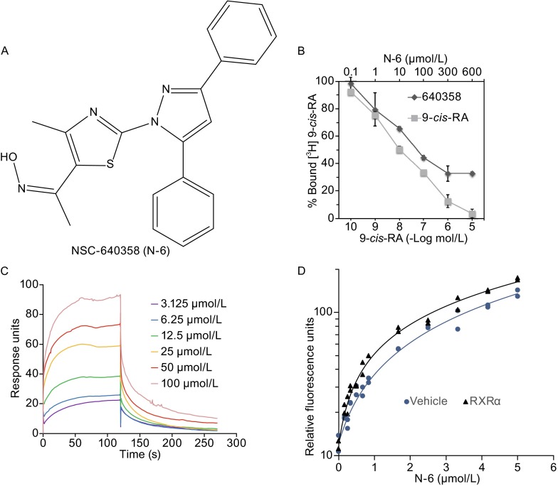
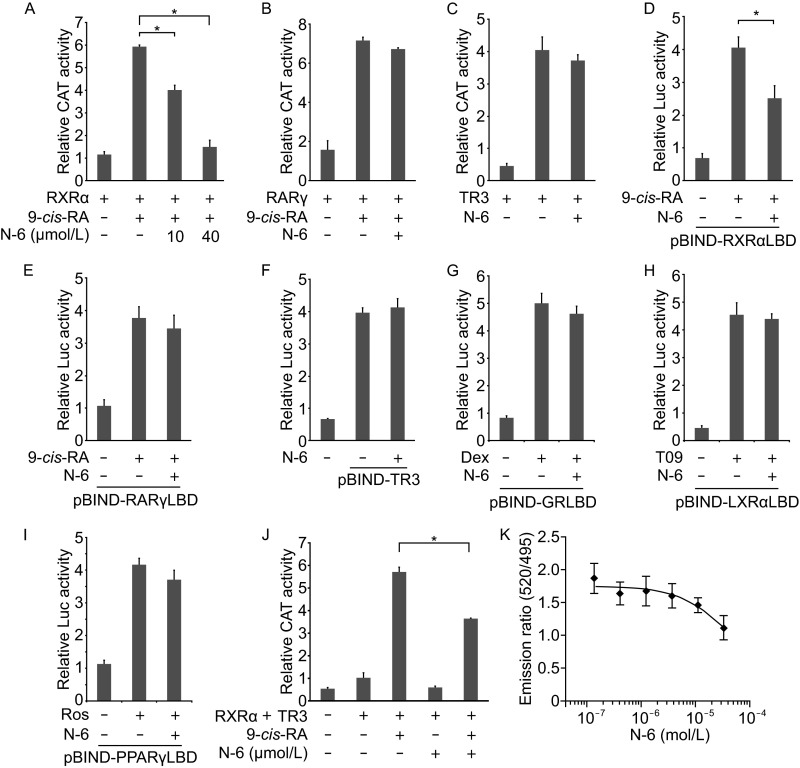
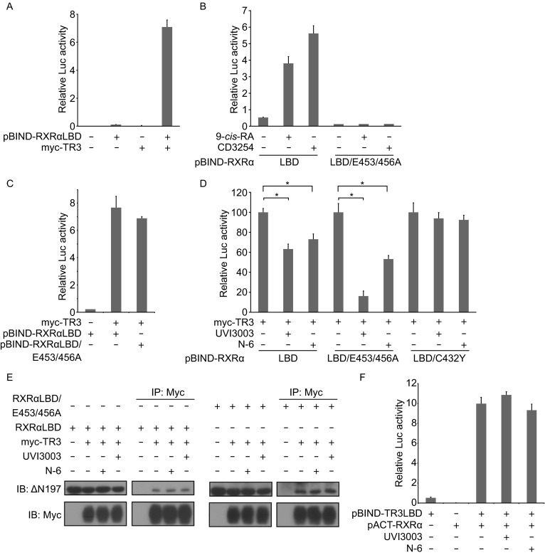
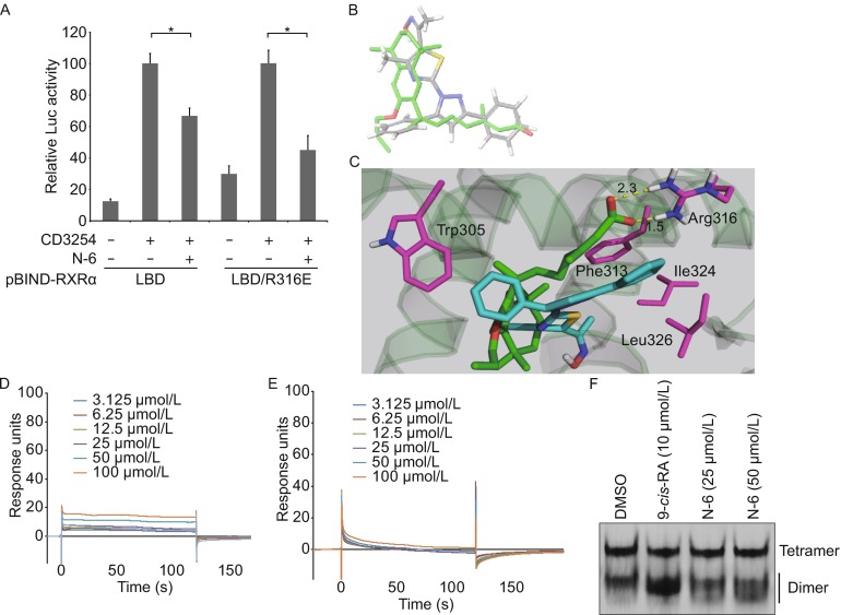
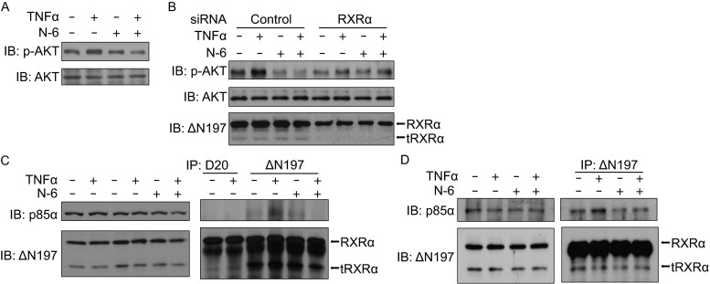
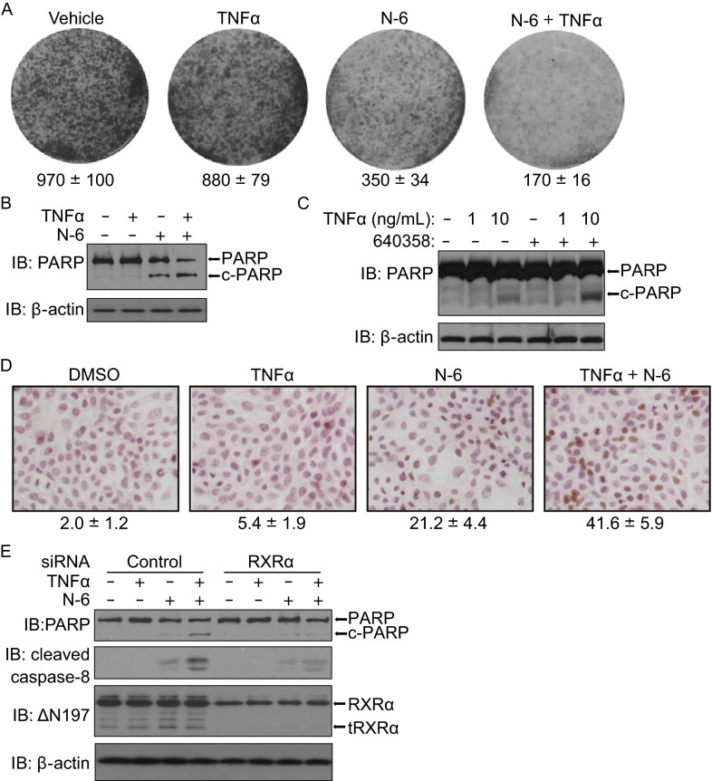
Retinoid X receptor α (RXRα) and its N-terminally truncated version tRXRα play important roles in tumorigenesis, while some RXRα ligands possess potent anti-cancer activities by targeting and modulating the tumorigenic effects of RXRα and tRXRα. Here we describe NSC-640358 (N-6), a thiazolyl-pyrazole derived compound, acts as a selective RXRα ligand to promote TNFα-mediated apoptosis of cancer cell. N-6 binds to RXRα and inhibits the transactivation of RXRα homodimer and RXRα/TR3 heterodimer. Using mutational analysis and computational study, we determine that Arg316 in RXRα, essential for 9-cis-retinoic acid binding and activating RXRα transactivation, is not required for antagonist effects of N-6, whereas Trp305 and Phe313 are crucial for N-6 binding to RXRα by forming extra π-π stacking interactions with N-6, indicating a distinct RXRα binding mode of N-6. N-6 inhibits TR3-stimulated transactivation of Gal4-DBD-RXRα-LBD by binding to the ligand binding pocket of RXRα-LBD, suggesting a strategy to regulate TR3 activity indirectly by using small molecules to target its interacting partner RXRα. For its physiological activities, we show that N-6 strongly inhibits tumor necrosis factor α (TNFα)-induced AKT activation and stimulates TNFα-mediated apoptosis in cancer cells in an RXRα/tRXRα dependent manner. The inhibition of TNFα-induced tRXRα/p85α complex formation by N-6 implies that N-6 targets tRXRα to inhibit TNFα-induced AKT activation and to induce cancer cell apoptosis. Together, our data illustrate a new RXRα ligand with a unique RXRα binding mode and the abilities to regulate TR3 activity indirectly and to induce TNFα-mediated cancer cell apoptosis by targeting RXRα/tRXRα.
Figures
Similar articles
-
Overview of the structure-based non-genomic effects of the nuclear receptor RXRα.Cell Mol Biol Lett. 2018 Aug 7;23:36. doi: 10.1186/s11658-018-0103-3. eCollection 2018. Cell Mol Biol Lett. 2018. PMID: 30093910 Free PMC article. Review.
-
Nitrostyrene Derivatives Act as RXRα Ligands to Inhibit TNFα Activation of NF-κB.Cancer Res. 2015 May 15;75(10):2049-60. doi: 10.1158/0008-5472.CAN-14-2435. Epub 2015 Mar 20. Cancer Res. 2015. PMID: 25795708 Free PMC article.
-
Antagonist effect of triptolide on AKT activation by truncated retinoid X receptor-alpha.PLoS One. 2012;7(4):e35722. doi: 10.1371/journal.pone.0035722. Epub 2012 Apr 24. PLoS One. 2012. PMID: 22545132 Free PMC article.
-
NSAID sulindac and its analog bind RXRalpha and inhibit RXRalpha-dependent AKT signaling.Cancer Cell. 2010 Jun 15;17(6):560-73. doi: 10.1016/j.ccr.2010.04.023. Cancer Cell. 2010. PMID: 20541701 Free PMC article.
-
Targeting truncated RXRα for cancer therapy.Acta Biochim Biophys Sin (Shanghai). 2016 Jan;48(1):49-59. doi: 10.1093/abbs/gmv104. Epub 2015 Oct 21. Acta Biochim Biophys Sin (Shanghai). 2016. PMID: 26494413 Free PMC article. Review.
Cited by
-
Implications of GPIIB-IIIA Integrin and Liver X Receptor in Platelet-Induced Compression of Ovarian Cancer Multi-Cellular Spheroids.Cancers (Basel). 2024 Oct 19;16(20):3533. doi: 10.3390/cancers16203533. Cancers (Basel). 2024. PMID: 39456628 Free PMC article.
-
Targeting mitochondria in cancer therapy could provide a basis for the selective anti-cancer activity.PLoS One. 2019 Mar 25;14(3):e0205623. doi: 10.1371/journal.pone.0205623. eCollection 2019. PLoS One. 2019. PMID: 30908483 Free PMC article.
-
Retinoid X Receptor Antagonists.Int J Mol Sci. 2018 Aug 10;19(8):2354. doi: 10.3390/ijms19082354. Int J Mol Sci. 2018. PMID: 30103423 Free PMC article. Review.
-
Inflammatory-Related P62 Triggers Malignant Transformation of Mesenchymal Stem Cells through the Cascade of CUDR-CTCF-IGFII-RAS Signaling.Mol Ther Nucleic Acids. 2018 Jun 1;11:367-381. doi: 10.1016/j.omtn.2018.03.002. Epub 2018 Mar 9. Mol Ther Nucleic Acids. 2018. Retraction in: Mol Ther Nucleic Acids. 2022 Dec 11;31:13. doi: 10.1016/j.omtn.2022.12.003. PMID: 29858072 Free PMC article. Retracted.
-
Overview of the structure-based non-genomic effects of the nuclear receptor RXRα.Cell Mol Biol Lett. 2018 Aug 7;23:36. doi: 10.1186/s11658-018-0103-3. eCollection 2018. Cell Mol Biol Lett. 2018. PMID: 30093910 Free PMC article. Review.
References
-
- Cao X, Liu W, Lin F, Li H, Kolluri SK, Lin B, Han YH, Dawson MI, Zhang XK. Retinoid X receptor regulates Nur77/TR3-dependent apoptosis [corrected] by modulating its nuclear export and mitochondrial targeting. Mol Cell Biol. 2004;24:9705–9725. doi: 10.1128/MCB.24.22.9705-9725.2004. - DOI - PMC - PubMed
Publication types
MeSH terms
Substances
Grants and funding
LinkOut - more resources
Full Text Sources
Other Literature Sources
Research Materials
Miscellaneous
