. 2015 Jul 22;282(1811):20151203.
doi: 10.1098/rspb.2015.1203.

Diffusion tensor imaging of dolphin brains reveals direct auditory pathway to temporal lobe

Affiliations

Diffusion tensor imaging of dolphin brains reveals direct auditory pathway to temporal lobe

Gregory S Berns et al. Proc Biol Sci. .

Abstract

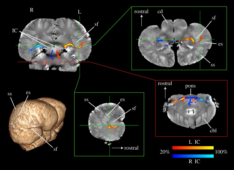
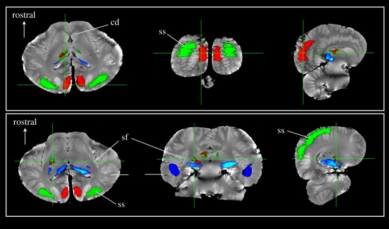
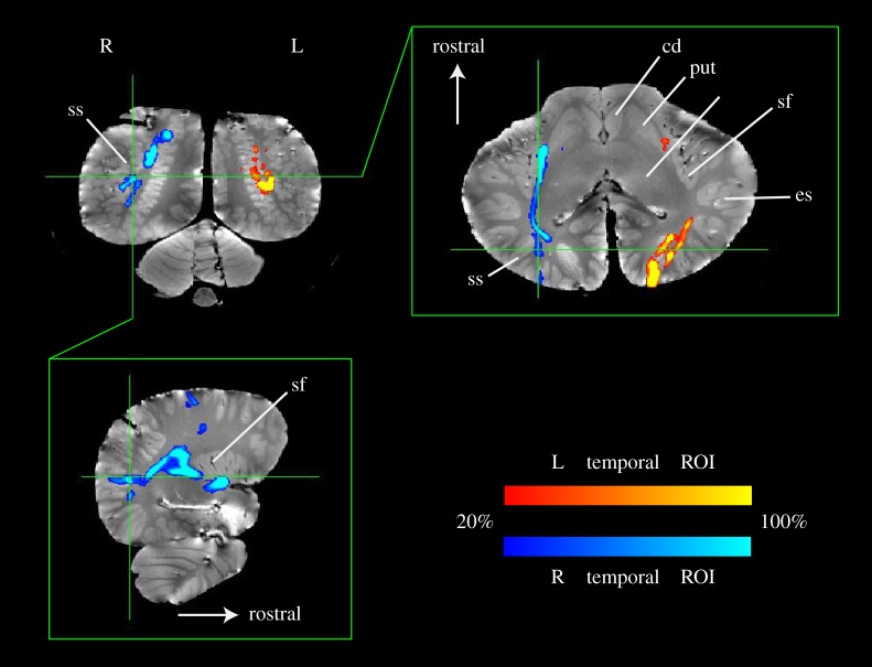
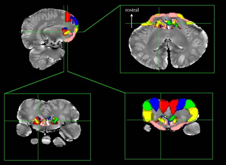
The brains of odontocetes (toothed whales) look grossly different from their terrestrial relatives. Because of their adaptation to the aquatic environment and their reliance on echolocation, the odontocetes' auditory system is both unique and crucial to their survival. Yet, scant data exist about the functional organization of the cetacean auditory system. A predominant hypothesis is that the primary auditory cortex lies in the suprasylvian gyrus along the vertex of the hemispheres, with this position induced by expansion of 'associative' regions in lateral and caudal directions. However, the precise location of the auditory cortex and its connections are still unknown. Here, we used a novel diffusion tensor imaging (DTI) sequence in archival post-mortem brains of a common dolphin (Delphinus delphis) and a pantropical dolphin (Stenella attenuata) to map their sensory and motor systems. Using thalamic parcellation based on traditionally defined regions for the primary visual (V1) and auditory cortex (A1), we found distinct regions of the thalamus connected to V1 and A1. But in addition to suprasylvian-A1, we report here, for the first time, the auditory cortex also exists in the temporal lobe, in a region near cetacean-A2 and possibly analogous to the primary auditory cortex in related terrestrial mammals (Artiodactyla). Using probabilistic tract tracing, we found a direct pathway from the inferior colliculus to the medial geniculate nucleus to the temporal lobe near the sylvian fissure. Our results demonstrate the feasibility of post-mortem DTI in archival specimens to answer basic questions in comparative neurobiology in a way that has not previously been possible and shows a link between the cetacean auditory system and those of terrestrial mammals. Given that fresh cetacean specimens are relatively rare, the ability to measure connectivity in archival specimens opens up a plethora of possibilities for investigating neuroanatomy in cetaceans and other species.

Keywords: auditory; diffusion tensor imaging; dolphin.


Figures

Figure 1.
Figure 1.
Probabilistic tractography from IC in a common dolphin brain (Delphinus delphis). Using seeds drawn on the left (red) and right (blue) inferior colliculi (IC, upper left), a tract to the temporal lobe was identified, which passed through the ventrocaudal thalamus and the medial geniculate nucleus (MGN). The tract colour indicates the percentage of streamlines passing through a voxel and is thresholded such that voxels in which less than 20% of the streamlines pass are not shown. Insets show transverse slices at the level of the MGN (upper right) and cochlear nuclei (lower right) which shows the decussation of the auditory tract and entrance of the auditory nerves, and a sagittal slice through the left sylvian fissure (lower middle). Three-dimensional rendering (lower left) shows major landmarks. IC, inferior colliculus; sf, sylvian fissure; ss, suprasylvian sulcus; es, ectosylvian sulcus; cd, caudate; cbl, cerebellum.
Figure 2.
Figure 2.
Thalamic parcellation of Delphinus delphis based on cortical regions. Three regions of interest were defined: (i) visual cortex (red); (ii) auditory cortex based on traditional boundaries in the suprasylvian gyrus (green) and (iii) auditory region temporal cortex based on IC tractography (blue). Seeds within the entire thalamus were traced to these regions and thresholded above 20 000 streamlines. Each row shows a set of slices that correspond to a single cursor location. The upper row is located posteriorly through the cortex while the lower row is located through thalamus. The thalamic parcellations to the three regions demonstrate that the temporal region is primarily connected to the ventrocaudal region, presumably near the medial geniculate nucleus. The visual and auditory regions overlap substantially in the thalamus and are located more dorsally and rostrally.
Figure 3.
Figure 3.
Probabilistic tractography from temporal ROIs in Delphinus delphis. Using seeds drawn on the left (red) and right (blue) temporal ROIs, tracts to the suprasylvian-A1 were identified. A ‘waypoint’ mask for suprasylvian-A1 was used to constrain the search space for tracts emanating from the temporal ROIs. The tract colour indicates the percentage of streamlines passing through a voxel and is thresholded such that voxels in which less than 20% of the streamlines pass are not shown.
Figure 4.
Figure 4.
Basal ganglia parcellation based on cortical regions in Delphinus delphis. Five regions were defined in the orbital (‘frontal’) lobes from the longitudinal fissure (red) progressing laterally and ventrally (blue, green and yellow) to the ventral orbital lobe (pink). Seeds within the entire basal ganglia were traced to these regions and thresholded above 20 000 streamlines. Bottom row shows two coronal slices through different parts of the basal ganglia. The dorsal portions of the basal ganglia are connected to the dorsal portions of the cortex (red, blue and green), while the ventral basal ganglia are connected to the ventrolateral (yellow) and ventral orbital lobe (pink).

Similar articles

Cited by

References

    1. Oelschläger HHA, Haas-Rioth M, Fung C, Ridgway SH, Knauth M. 2008. Morphology and evolutionary biology of the dolphin (Delphinus sp.) brain—MR imaging and conventional histology. Brain Behav. Evol. 71, 68–86. (10.1159/000110495) - DOI - PubMed
    1. Oelschlager HHA, Ridgway SH, Knauth M. 2010. Cetacean brain evolution: dwarf sperm whale (Kogia sima) and common dolphin (Delphinus delphis)—an investigation with high-resolution 3D MRI. Brain Behav. Evol. 75, 33–62. (10.1159/000293601) - DOI - PubMed
    1. Marino L, Sudheimer KD, Pabst DA, McLellan WA, Filsoof D, Johnson JI. 2002. Neuroanatomy of the common dolphin (Delphinus delphis) as revealed by magnetic resonance imaging. Anat. Rec. 268, 411–429. (10.1002/ar.10181) - DOI - PubMed
    1. Montie EW, Ketten DR, Schneider G, Marino L, Touhey KE, Hahn ME. 2007. Neuroanatomy of the subadult and fetal brain of the Atlantic white-sided dolphin (Lagenorhynchus acutus) from in situ magnetic resonance images. Anat. Rec. 290, 1459–1479. (10.1002/ar.20612) - DOI - PubMed
    1. Marino L, Murphy TL, DeWeerd AL, Morris JA, Ridgway SH, Fobbs AJ, Humblot N, Johnson JI. 2001. Anatomy and three-dimensional reconstructions of the brain of a white whale (Delphinapterus leucas) from magnetic resonance images (MRI). Anat. Rec. 262, 429–439. (10.1002/ar.1051) - DOI - PubMed

Publication types

MeSH terms