. 2015 Jul 8;35(27):9879-88.
doi: 10.1523/JNEUROSCI.1239-15.2015.

Neuronal Differentiation in Schwann Cell Lineage Underlies Postnatal Neurogenesis in the Enteric Nervous System

Affiliations

Neuronal Differentiation in Schwann Cell Lineage Underlies Postnatal Neurogenesis in the Enteric Nervous System

Toshihiro Uesaka et al. J Neurosci. .

Abstract

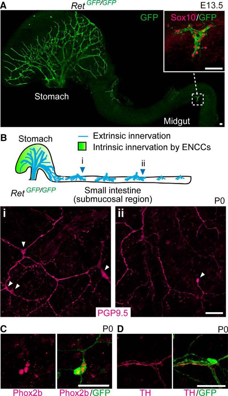
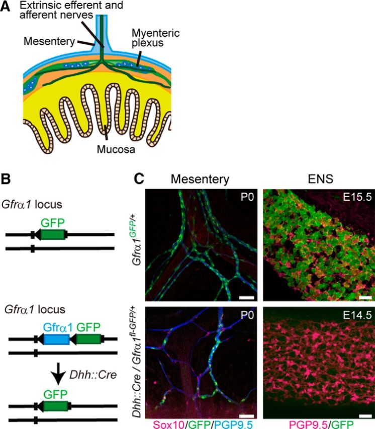
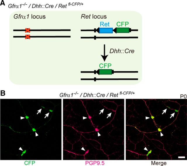
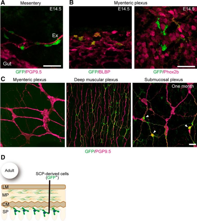
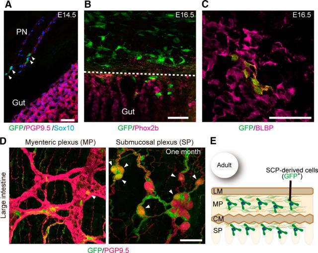
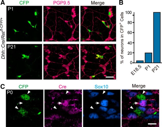
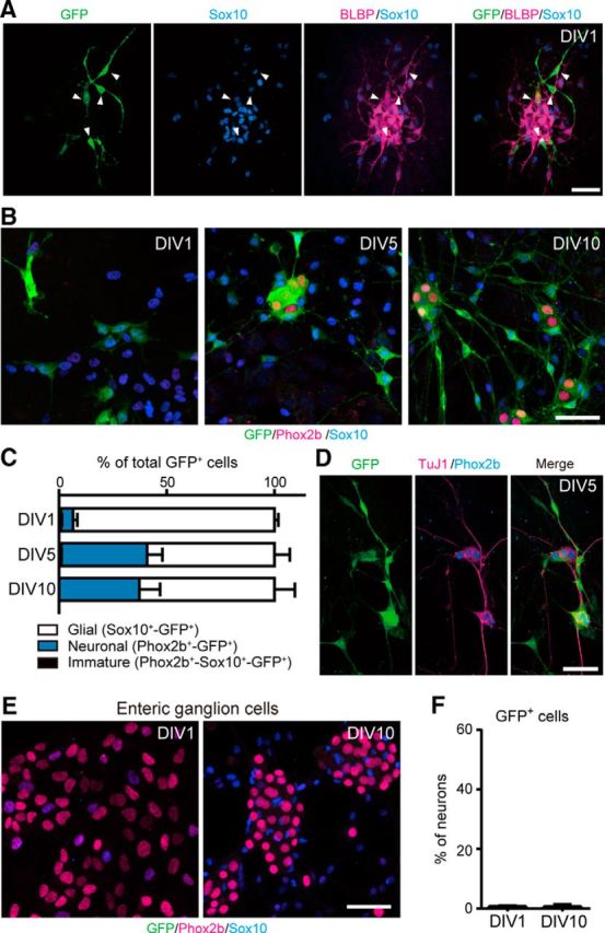
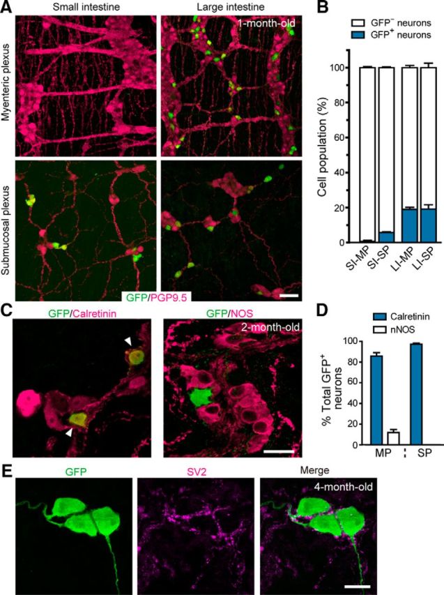
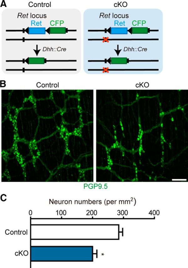
Elucidation of the cellular identity of neuronal precursors provides mechanistic insights into the development and pathophysiology of the nervous system. In the enteric nervous system (ENS), neurogenesis persists from midgestation to the postnatal period. Cellular mechanism underlying the long-term neurogenesis in the ENS has remained unclear. Using genetic fate mapping in mice, we show here that a subset of Schwann cell precursors (SCPs), which invades the gut alongside the extrinsic nerves, adopts a neuronal fate in the postnatal period and contributes to the ENS. We found SCP-derived neurogenesis in the submucosal region of the small intestine in the absence of vagal neural crest-derived ENS precursors. Under physiological conditions, SCPs comprised up to 20% of enteric neurons in the large intestine and gave rise mainly to restricted neuronal subtypes, calretinin-expressing neurons. Genetic ablation of Ret, the signaling receptor for glial cell line-derived neurotrophic factor, in SCPs caused colonic oligoganglionosis, indicating that SCP-derived neurogenesis is essential to ENS integrity. Identification of Schwann cells as a physiological neurogenic source provides novel insight into the development and disorders of neural crest-derived tissues.

Significance statement: Elucidating the cellular identity of neuronal precursors provides novel insights into development and function of the nervous system. The enteric nervous system (ENS) is innervated richly by extrinsic nerve fibers, but little is known about the significance of extrinsic innervation to the structural integrity of the ENS. This report reveals that a subset of Schwann cell precursors (SCPs), which invades the gut alongside the extrinsic nerves, adopts a neuronal fate and differentiates into specific neuronal subtypes. SCP-specific ablation of the Ret gene leads to colonic oligoganglionosis, demonstrating a crucial role of SCP-derived neurogenesis in ENS development. Cross-lineage differentiation capacity in SCPs suggests their potential involvement in the development and pathology of a wide variety of neural crest-derived cell types.

Keywords: RET; Schwann cells; enteric nervous system; neural crest cells; neurogenesis; oligoganglionosis.


Figures

Figure 1.
Figure 1.
Enteric neurogenesis occurs without vagal ENCCs. A, Whole-mount GFP (green) staining of the gastrointestinal tract from E13.5 Ret-deficient (RetGFP/GFP) embryos (n = 3). Higher magnification (inset) shows the presence of Sox10+ SCPs along the extrinsic nerve bundles. B, Top, Schematic diagram illustrating the stomach and the locations of extrinsic nerve fibers. Bottom, Representative images of the submucosal region in RetGFP/GFP small intestine (i, ii), showing expression of PGP9.5 (magenta; white arrowheads in i and ii, n = 3). C, Whole-mount GFP (green) and Phox2b (magenta) staining of the submucosal region in RetGFP/GFP duodenum (n = 3). D, Submucosal neurons in RetGFP/GFP mice (n = 3) associated with tyrosine hydroxylase-positive (TH+) sympathetic nerve fibers (magenta). Scale bars, 50 μm.
Figure 2.
Figure 2.
SCPs are labeled selectively by GFP in Dhh::Cre/Gfrα1fl-GFP mice. A, Schematic diagram showing extrinsic nerve fibers projecting to the gut at E14.5. B, Schematic representation of Gfrα1–GFP knock-in allele for the Gfrα1fl–GFP reporter (top) and genetic tracing of SCPs by Dhh::Cre and Gfrα1fl–GFP reporter line (bottom). C, Gfrα1 promoter drives GFP expression in both SCPs along the extrinsic nerves (top left) and ENS cells (top right) in Gfrα1GFP/+ mice (P0, n = 3; E15.5, n = 3). Dhh::Cre-mediated GFP labeling was detected in SCPs (Sox10+) within the mesentery (bottom left, n = 3 mice), whereas GFP signals were not detected in vagal NC-derived enteric neurons (PGP9.5+) at E14.5 (bottom right, n = 3). Scale bars, 50 μm.
Figure 3.
Figure 3.
Enteric neurons in Gfrα1-deficient small intestine are labeled by the Dhh::Cre driver. A, Schematic showing genetic tracing of SCPs in Gfrα1-deficient mice by the Dhh::Cre driver. B, Confocal images of Gfrα1-deficient small intestine (Dhh::Cre/Retfl-CFP/+/Gfrα1−/−, n = 3) showing the presence of GFP+–PGP9.5+ neurons (arrowheads) in the submucosal region. Small arrows depict GFP+–PGP9.5 cells. Scale bar, 50 μm.
Figure 4.
Figure 4.
Pattern of colonization by SCPs in the small intestine. A, Representative image showing the presence of GFP+ cells along the extrinsic nerve (Ex) in the mesentery of E14.5 Dhh::Cre/Gfrα1fl-GFP/+ fetuses (n = 3). B, Whole-mount GFP (green) and BLBP (magenta, left) or Phox2b (right) staining of Dhh::Cre/Gfrα1fl-GFP/+ gut (n = 3) revealing that GFP+ cells invading the myenteric plexus at E14.5 express BLBP (a glial marker) but not Phox2b (a marker for progenitor and neuron). C, Representative images of whole-mount GFP (green) and PGP9.5 (magenta) staining of the myenteric, deep muscular, and submucosal plexuses of the small intestine of Dhh::Cre/Gfrα1fl–GFP mice aged 1 month (n = 4). SCPs are barely detectable in the myenteric ganglia (left) but are abundant in the deep muscular plexus (middle). A few GFP-labeled neurons (arrowheads) are present in the submucosal plexus of the small intestine (right). D, Schematic showing the colonization pattern of SCP-derived cells in adult small intestine. Scale bars: A, B, 50 μm; C, 20 μm. LM, Longitudinal muscle layers; MP, myenteric plexus; CM, circular muscle layers; SP, submucosal plexus.
Figure 5.
Figure 5.
SCPs colonize both the myenteric and submucosal plexuses in the large intestine. A, Comparison of colonization pattern between sacral ENCCs and SCPs in the distal colon. SCPs (green, arrowheads), which were labeled by the Dhh::Cre driver, were detected only along the pelvic nerves (PN) in Dhh::Cre/Gfrα1fl–GFP embryos (E14.5, n = 3). PGP9.5 staining (red) shows intrinsic ENCC innervation (gut) and the pelvic nerve. Sacral ENCCs were not labeled by the Dhh::Cre driver. B, A representative image showing migration of SCPs from the pelvic nerve to the wall of distal hindgut at E16.5 (n = 3). SCPs in the myenteric plexus did not express Phox2b during this period. C, Whole-mount GFP (green) and BLBP (magenta) staining of the large intestine of Dhh::Cre/Gfrα1fl-GFP/+ mice (n = 3), revealing that GFP+ cells invading the myenteric plexus at E16.5 express BLBP. D, Localization of SCPs (GFP+, green) in the myenteric and submucosal plexuses of the large intestine (n = 3) at 1 month. Neuronal cells were visualized by PGP9.5 (magenta) antibodies. Arrowheads indicate GFP+–PGP9.5+ neurons. E, Schematic showing the colonization pattern of SCP-derived cells in adult large intestine. Scale bars, 50 μm. LM, Longitudinal muscle layers; MP, myenteric plexus; CM, circular muscle layers; SP, submucosal plexus.
Figure 6.
Figure 6.
Postnatal neuronal differentiation of SCPs. A, Representative images of whole-mount GFP (green) and PGP9.5 (magenta) staining of Dhh::Cre/Retfl-CFP/+ submucosal plexus of the small intestine at P1 and P21. B, Quantification of the fraction of GFP+–PGP9.5+ double-positive neurons within the GFP+ cells in the small intestine at E18.5, P1, and P21 (n = 3 animals per group). C, Representative images of whole-mount CFP (Ret+) and Cre recombinase staining of Dhh::Cre/Retfl-CFP/+ submucosal plexus of the small intestine (n = 3). During the period of neuronal differentiation (indicated by the emergence of Ret), no Cre recombinase was detected in any of Ret+ cells. Arrow indicates Cre recombinase detected in Sox10+ glial cells. Scale bars: A, 50 μm; C, 20 μm.
Figure 7.
Figure 7.
Extrinsic nerve-associated SCPs display neurogenic potential in vitro. A, GFP-labeled cells isolated from the mesentery of E16.5 Dhh::Cre/Gfrα1fl–GFP embryos expressed the glial cell markers Sox10 (blue) and BLBP (magenta) 1 d after plating [1 d in vitro (DIV1)]. B, Representative images of cultured cells stained with GFP (green), Sox10 (blue), and Phox2b (magenta) antibodies at 1, 5, and 10 d after plating. At DIV1, most of GFP+ cells expressed Sox10. At the date of plating, there were very few, if any, GFP+ cells expressing Phox2b, a marker for enteric neurons. After 5 d in culture, a substantial population of GFP+ cells acquired Phox2b expression (DIV5 and DIV10). C, Quantification of the fraction of neurons, glia, and immature cells in GFP+ cell populations of DIV1, DIV5, and DIV10 cultures (n = 4 experiments; error bars indicate SEM). D, Immunostaining by the pan-neuronal marker TuJ1 (magenta) confirmed that GFP+–Phox2b+ cells are neurons. E, ENCCs isolated from E16.5 Dhh::Cre/Gfrα1fl–GFP midgut did not express Dhh::Cre during 10 d incubations. F, Quantification of a GFP+ neuronal population in ENCC-derived neurons of DIV1 and DIV10 cultures (n = 3 experiments; error bars indicate SEM). Scale bars, 50 μm.
Figure 8.
Figure 8.
Contribution of SCP-derived neurons to the ENS in the postnatal period. A, Representative images of the myenteric and submucosal plexus (top and bottom panels) in the small and large intestine (left and right) of Dhh::Cre/Retfl-CFP/+ mice (1-month-old), visualizing SCP-derived neurons (CFP, green) and all enteric neurons (PGP9.5, magenta). B, Contribution of SCPs to enteric neurons (blue bars) calculated by dividing the numbers of SCP-derived neurons (CFP+) by those of all enteric neurons (PGP9.5+). SI, Small intestine; LI, large intestine; MP, myenteric plexus; SP, submucosal plexus (n = 3 animals; error bars represent SEM). C, Representative images of myenteric plexus in the large intestine stained with antibodies to GFP, calretinin, and NOS. D, Percentage of calretinin+ (blue bars) and NOS+ neurons (white bars) in SCP-derived neurons of the myenteric plexus (MP) and submucosal plexus (SP) (n = 3 animals; error bars represent SEM). E, Representative images showing presence of SV2 around the cell bodies and dendrites of GFP+ enteric neurons in 4-month-old mice (n = 3). Scale bars: A, 50 μm; C, E, 20 μm.
Figure 9.
Figure 9.
Ret inactivation in SCPs causes hypoganglionosis in the terminal colon. A, Schematic showing conditional inactivation of the Ret gene in SCPs by Dhh::Cre. The Ret locus of the control and cKO animals harbors wild-type and null (orange cross) alleles, respectively, in addition to the Retfl–CFP reporter allele. B, Representative images of whole-mount PGP9.5 staining of the distal colon of control and cKO mice aged 4 months. C, Quantification of enteric neurons in the terminal colon of control and cKO mice at 4 months. Significant differences detected by t test with Welch's correction (n = 3 mice from 3 litters; error bars represent SEM; F(2,2) = 1.263, *p = 0.0204). Scale bars, 50 μm.

Similar articles

Cited by

References

    1. Adameyko I, Lallemend F, Aquino JB, Pereira JA, Topilko P, Müller T, Fritz N, Beljajeva A, Mochii M, Liste I, Usoskin D, Suter U, Birchmeier C, Ernfors P. Schwann cell precursors from nerve innervation are a cellular origin of melanocytes in skin. Cell. 2009;139:366–379. doi: 10.1016/j.cell.2009.07.049. - DOI - PubMed
    1. Anderson RB, Stewart AL, Young HM. Phenotypes of neural-crest-derived cells in vagal and sacral pathways. Cell Tissue Res. 2006;323:11–25. doi: 10.1007/s00441-005-0047-6. - DOI - PubMed
    1. Ayer-Le Lievre CS, Le Douarin NM. The early development of cranial sensory ganglia and the potentialities of their component cells studied in quail-chick chimeras. Dev Biol. 1982;94:291–310. doi: 10.1016/0012-1606(82)90349-9. - DOI - PubMed
    1. Bitgood MJ, McMahon AP. Hedgehog and Bmp genes are coexpressed at many diverse sites of cell-cell interaction in the mouse embryo. Dev Biol. 1995;172:126–138. doi: 10.1006/dbio.1995.0010. - DOI - PubMed
    1. Branchek TA, Gershon MD. Time course of expression of neuropeptide Y, calcitonin gene-related peptide, and NADPH diaphorase activity in neurons of the developing murine bowel and the appearance of 5-hydroxytryptamine in mucosal enterochromaffin cells. J Comp Neurol. 1989;285:262–273. doi: 10.1002/cne.902850208. - DOI - PubMed

Publication types

MeSH terms