

Catecholaminergic neurons projecting to the paraventricular nucleus of the hypothalamus are essential for cardiorespiratory adjustments to hypoxia
- PMID: 26157062
- PMCID: PMC4666929
- DOI: 10.1152/ajpregu.00540.2014
Catecholaminergic neurons projecting to the paraventricular nucleus of the hypothalamus are essential for cardiorespiratory adjustments to hypoxia
Abstract
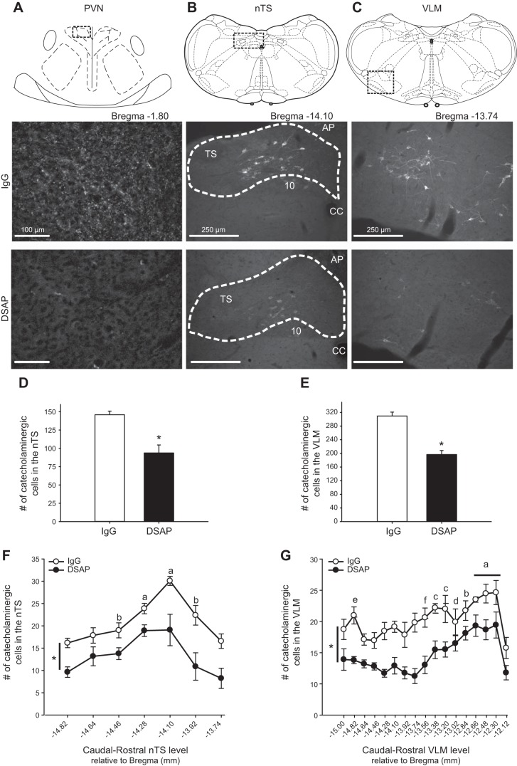
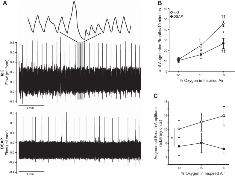
Brainstem catecholamine neurons modulate sensory information and participate in control of cardiorespiratory function. These neurons have multiple projections, including to the paraventricular nucleus (PVN), which contributes to cardiorespiratory and neuroendocrine responses to hypoxia. We have shown that PVN-projecting catecholaminergic neurons are activated by hypoxia, but the function of these neurons is not known. To test the hypothesis that PVN-projecting catecholamine neurons participate in responses to respiratory challenges, we injected IgG saporin (control; n = 6) or anti-dopamine β-hydroxylase saporin (DSAP; n = 6) into the PVN to retrogradely lesion catecholamine neurons projecting to the PVN. After 2 wk, respiratory measurements (plethysmography) were made in awake rats during normoxia, increasing intensities of hypoxia (12, 10, and 8% O2) and hypercapnia (5% CO2-95% O2). DSAP decreased the number of tyrosine hydroxylase-immunoreactive terminals in PVN and cells counted in ventrolateral medulla (VLM; -37%) and nucleus tractus solitarii (nTS; -36%). DSAP produced a small but significant decrease in respiratory rate at baseline (during normoxia) and at all intensities of hypoxia. Tidal volume and minute ventilation (VE) index also were impaired at higher hypoxic intensities (10-8% O2; e.g., VE at 8% O2: IgG = 181 ± 22, DSAP = 91 ± 4 arbitrary units). Depressed ventilation in DSAP rats was associated with significantly lower arterial O2 saturation at all hypoxic intensities. PVN DSAP also reduced ventilatory responses to 5% CO2 (VE: IgG = 176 ± 21 and DSAP = 84 ± 5 arbitrary units). Data indicate that catecholamine neurons projecting to the PVN are important for peripheral and central chemoreflex respiratory responses and for maintenance of arterial oxygen levels during hypoxic stimuli.
Keywords: anti-dopamine β-hydroxylase saporin; blood pressure; brainstem; chemoreflex; ventilation.
Copyright © 2015 the American Physiological Society.
Figures


Similar articles
-
Hypoxia activates nucleus tractus solitarii neurons projecting to the paraventricular nucleus of the hypothalamus.Am J Physiol Regul Integr Comp Physiol. 2012 May 15;302(10):R1219-32. doi: 10.1152/ajpregu.00028.2012. Epub 2012 Mar 7. Am J Physiol Regul Integr Comp Physiol. 2012. PMID: 22403798 Free PMC article.
-
Acute systemic hypoxia activates hypothalamic paraventricular nucleus-projecting catecholaminergic neurons in the caudal ventrolateral medulla.Am J Physiol Regul Integr Comp Physiol. 2013 Nov 15;305(10):R1112-23. doi: 10.1152/ajpregu.00280.2013. Epub 2013 Sep 18. Am J Physiol Regul Integr Comp Physiol. 2013. PMID: 24049118 Free PMC article.
-
Paraventricular nucleus projections to the nucleus tractus solitarii are essential for full expression of hypoxia-induced peripheral chemoreflex responses.J Physiol. 2023 Oct;601(19):4309-4336. doi: 10.1113/JP284907. Epub 2023 Aug 26. J Physiol. 2023. PMID: 37632733
-
Branching projections of catecholaminergic brainstem neurons to the paraventricular hypothalamic nucleus and the central nucleus of the amygdala in the rat.Brain Res. 1993 Apr 23;609(1-2):81-92. doi: 10.1016/0006-8993(93)90858-k. Brain Res. 1993. PMID: 8099526
-
Modulation of cardiorespiratory function mediated by the paraventricular nucleus.Respir Physiol Neurobiol. 2010 Nov 30;174(1-2):55-64. doi: 10.1016/j.resp.2010.08.001. Epub 2010 Aug 11. Respir Physiol Neurobiol. 2010. PMID: 20708107 Free PMC article. Review.
Cited by
-
Unilateral vagotomy alters astrocyte and microglial morphology in the nucleus tractus solitarii of the rat.Am J Physiol Regul Integr Comp Physiol. 2021 Jun 1;320(6):R945-R959. doi: 10.1152/ajpregu.00019.2021. Epub 2021 May 12. Am J Physiol Regul Integr Comp Physiol. 2021. PMID: 33978480 Free PMC article.
-
Hydrogen peroxide inhibits neurons in the paraventricular nucleus of the hypothalamus via potassium channel activation.Am J Physiol Regul Integr Comp Physiol. 2019 Jul 1;317(1):R121-R133. doi: 10.1152/ajpregu.00054.2019. Epub 2019 May 1. Am J Physiol Regul Integr Comp Physiol. 2019. PMID: 31042419 Free PMC article.
-
Central AT1 receptor signaling by circulating angiotensin II is permissive to acute intermittent hypoxia-induced sympathetic neuroplasticity.J Appl Physiol (1985). 2020 May 1;128(5):1329-1337. doi: 10.1152/japplphysiol.00094.2020. Epub 2020 Apr 2. J Appl Physiol (1985). 2020. PMID: 32240022 Free PMC article.
-
Neural Control of Blood Pressure in Chronic Intermittent Hypoxia.Curr Hypertens Rep. 2016 Mar;18(3):19. doi: 10.1007/s11906-016-0627-8. Curr Hypertens Rep. 2016. PMID: 26838032 Free PMC article. Review.
-
Medullary Noradrenergic Neurons Mediate Hemodynamic Responses to Osmotic and Volume Challenges.Front Physiol. 2021 Apr 23;12:649535. doi: 10.3389/fphys.2021.649535. eCollection 2021. Front Physiol. 2021. PMID: 33967822 Free PMC article.
References
-
- Andresen MC, Kunze DL. Nucleus tractus solitarius - gateway to neural circulatory control. Annu Rev Physiol 56: 93–116, 1994. - PubMed
-
- Berquin P, Bodineau L, Gros F, Larnicol N. Brainstem and hypothalamic areas involved in respiratory chemoreflexes: a Fos study in adult rats. Brain Res 857: 30–40, 2000. - PubMed
Publication types
MeSH terms
Substances
Grants and funding
LinkOut - more resources
Full Text Sources
Other Literature Sources