. 2015 Sep;169(1):512-29.
doi: 10.1104/pp.15.00251. Epub 2015 Jul 8.

Extra-Large G Proteins Expand the Repertoire of Subunits in Arabidopsis Heterotrimeric G Protein Signaling

Affiliations

Extra-Large G Proteins Expand the Repertoire of Subunits in Arabidopsis Heterotrimeric G Protein Signaling

David Chakravorty et al. Plant Physiol. 2015 Sep.

Abstract

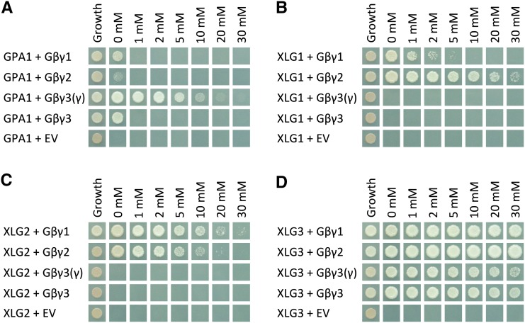
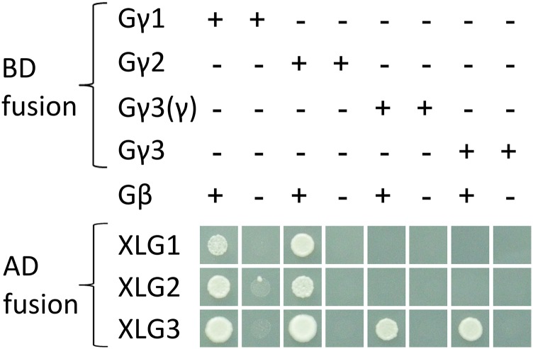
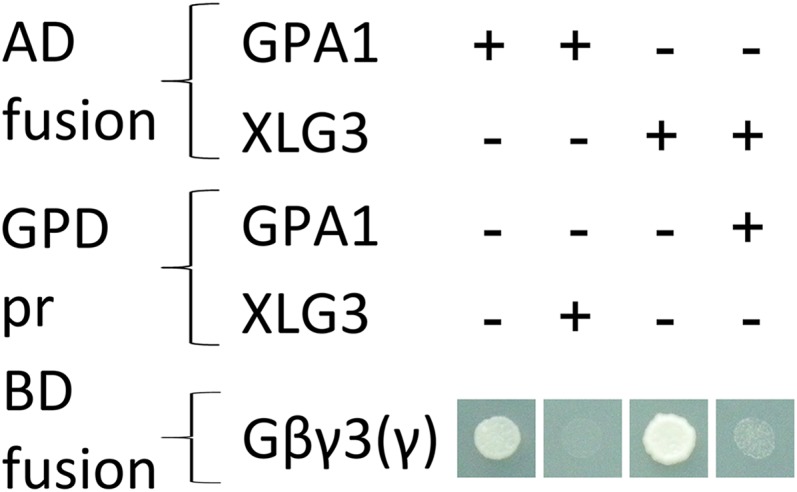
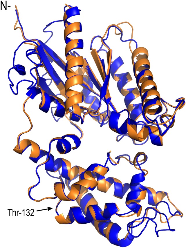
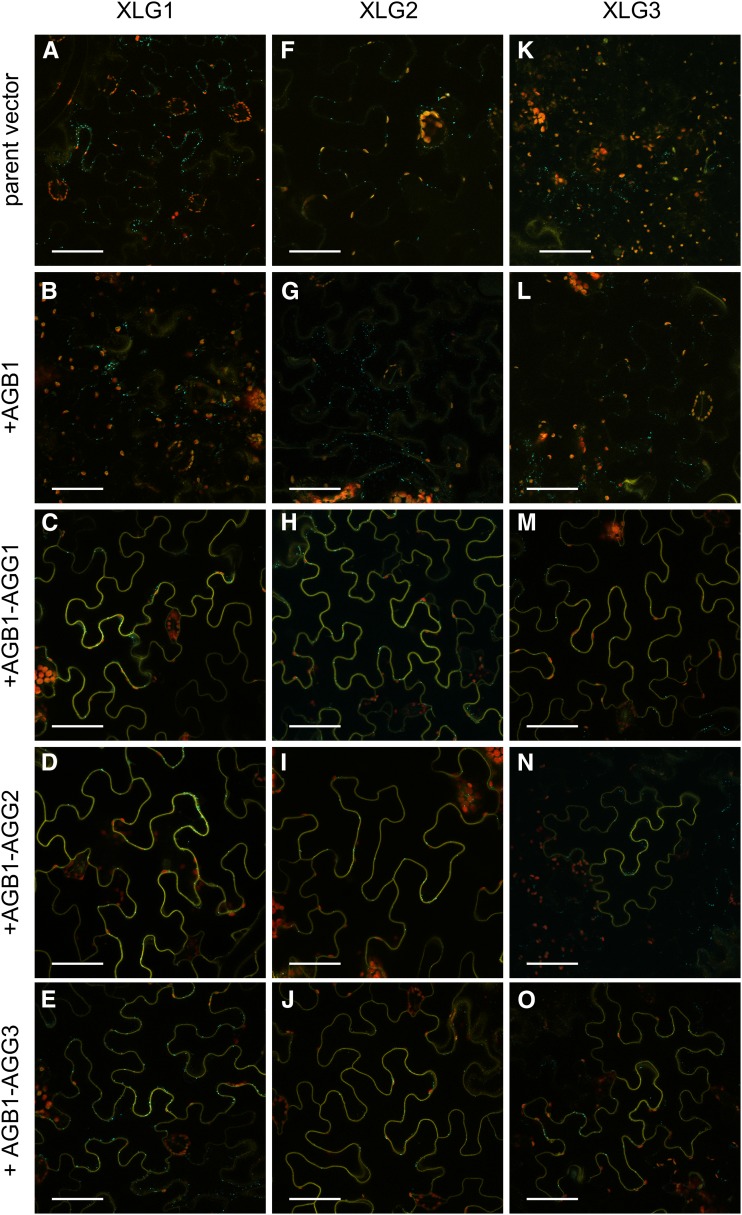
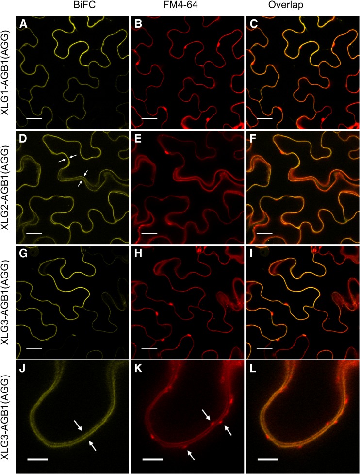
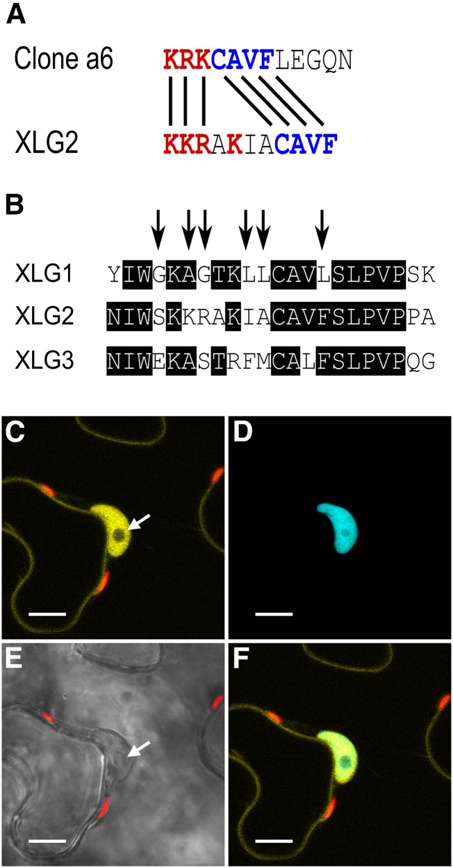
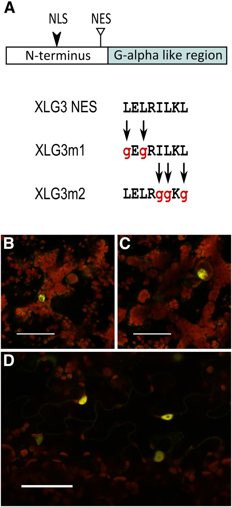
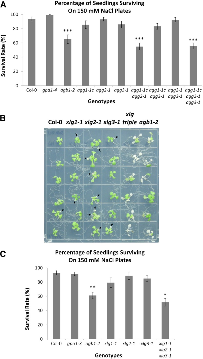
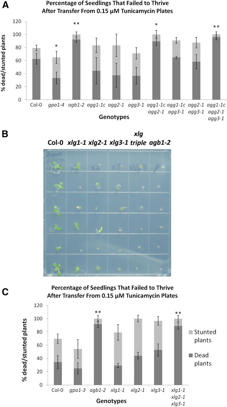
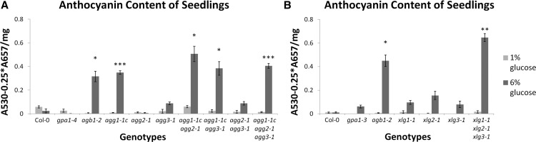
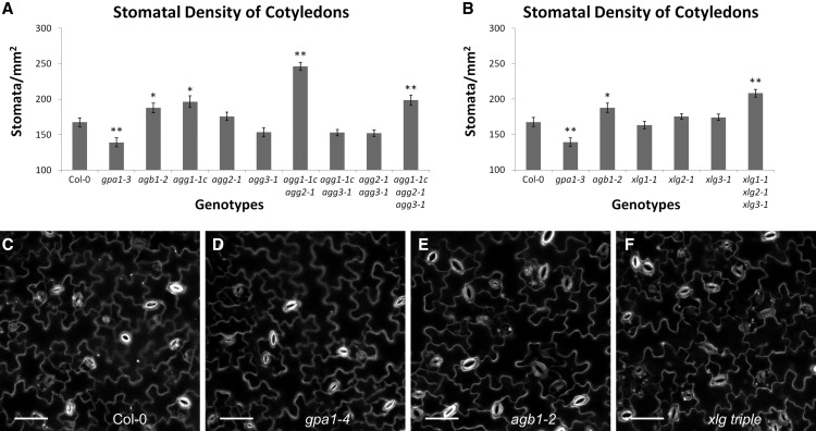
Heterotrimeric G proteins, consisting of Gα, Gβ, and Gγ subunits, are a conserved signal transduction mechanism in eukaryotes. However, G protein subunit numbers in diploid plant genomes are greatly reduced as compared with animals and do not correlate with the diversity of functions and phenotypes in which heterotrimeric G proteins have been implicated. In addition to GPA1, the sole canonical Arabidopsis (Arabidopsis thaliana) Gα subunit, Arabidopsis has three related proteins: the extra-large GTP-binding proteins XLG1, XLG2, and XLG3. We demonstrate that the XLGs can bind Gβγ dimers (AGB1 plus a Gγ subunit: AGG1, AGG2, or AGG3) with differing specificity in yeast (Saccharomyces cerevisiae) three-hybrid assays. Our in silico structural analysis shows that XLG3 aligns closely to the crystal structure of GPA1, and XLG3 also competes with GPA1 for Gβγ binding in yeast. We observed interaction of the XLGs with all three Gβγ dimers at the plasma membrane in planta by bimolecular fluorescence complementation. Bioinformatic and localization studies identified and confirmed nuclear localization signals in XLG2 and XLG3 and a nuclear export signal in XLG3, which may facilitate intracellular shuttling. We found that tunicamycin, salt, and glucose hypersensitivity and increased stomatal density are agb1-specific phenotypes that are not observed in gpa1 mutants but are recapitulated in xlg mutants. Thus, XLG-Gβγ heterotrimers provide additional signaling modalities for tuning plant G protein responses and increase the repertoire of G protein heterotrimer combinations from three to 12. The potential for signal partitioning and competition between the XLGs and GPA1 is a new paradigm for plant-specific cell signaling.


Figures

Figure 1.
Figure 1.
Yeast three-hybrid assays demonstrate the specificity of GPA1 for AGB1/AGG3, XLG1 and XLG2 for AGB1/AGG1 and AGB1/AGG2, and XLG3 for all three Gβγ dimers. Yeast three-hybrid assays tested the interactions between GPA1 (A), XLG1 (B), XLG2 (C), and XLG3 (D; GAL4 activation domain fusions) with the AGB1/AGG1 (Gβγ1), AGB1/AGG2 (Gβγ2), and AGB1/AGG3 (Gβγ3) Gβγ dimers (Gγ fused to the GAL4 binding domain, with Gβ [AGB1] as the bridge protein). A truncated γ3(γ) was also tested, consisting of only the Gγ-like domain of AGG3 (residues 1–112), lacking the implicated transmembrane domain. Yeast growth on synthetic complete (SC)-Trp-Leu confirmed transformation and cell viability. Interactions were assayed on SC-Trp-Leu-Met-His supplemented with the indicated concentrations of 3-AT (0–30 mm). EV, Empty vector.
Figure 2.
Figure 2.
The interaction of XLGs with Gγ subunits is AGB1 dependent. Yeast three-hybrid assays tested the interactions of GPA1, XLG1, XLG2, and XLG3 with the AGG1 (Gγ1), AGG2 (Gγ2), and AGG3 (Gγ3) subunits. Interactions were assayed in the presence and absence of AGB1. A truncated γ3(γ) subunit was also included, which includes only the Gγ-like domain of AGG3 (residues 1–112). All interactions were assayed by growth on SC-Trp-Leu-Met-His supplemented with 2 mm 3-AT. Two millimolars of 3-AT was used because the pBridge-AGG1 construct exhibited autoactivation (i.e. growth in combination with the empty vector) on SC-Trp-Leu-Met-His supplemented with 0 or 1 mm 3-AT.
Figure 3.
Figure 3.
GPA1 and XLG3 compete with each other for binding of AGB1/AGG3 in yeast. Yeast three-hybrid competition assays tested the interaction between GPA1 and AGB1/AGG3(γ) [Gβγ3(γ)] with or without additional expression of XLG3 and the interaction between XLG3 and AGB1/AGG3(γ) [Gβγ3(γ)] with or without additional expression of GPA1. The fourth protein (XLG3 or GPA1, respectively) was expressed under the control of the strong GPD promoter (pr). All interactions were assayed by growth on SC-Trp-Leu-Met-His supplemented with 2 mm 3-AT.
Figure 4.
Figure 4.
The XLG3 Gα-like region aligns closely to the crystal structure of GPA1. Superimposition of computationally derived XLG3 structural features on the empirically derived GPA1 crystal structure demonstrates their shared three-dimensional characteristics. Blue indicates the XLG3 Gα-like region, orange indicates the GPA1 Gα-like region, and N- indicates the N terminus of the Gα regions. Thr-132 marks the NVen210 insertion site in the αB-αC loop of GPA1 used for BiFC (Gookin and Assmann, 2014): each of the XLGs was modified at the analogous residue.
Figure 5.
Figure 5.
XLG1, XLG2, and XLG3 interact with AGB1 at the plasma membrane in an AGG-dependent manner. In all assays, positive transformation is confirmed by Golgi-localized mTurquoise2 (mTq2) fluorescence from the pDOE XT-Golgi-mTq2 marker. A, The XLG1L-CVen210 parent vector does not produce nonspecific BiFC signal. B, The XLG1L-CVen210:AGB1 construct shows zero to near-zero signal in the absence of a coexpressed Gγ subunit. C, The XLG1L-CVen210:AGB1 construct produces BiFC signal in the presence of coexpressed AGG1. D, The XLG1L-CVen210:AGB1 construct produces BiFC signal in the presence of coexpressed AGG2. E, The XLG1L-CVen210:AGB1 construct produces BiFC signal in the presence of coexpressed AGG3. F, The XLG2L-CVen210 parent vector does not produce nonspecific BiFC signal. G, The XLG2L-CVen210:AGB1 construct shows zero to near-zero signal in the absence of a coexpressed Gγ subunit. H, The XLG2L-CVen210:AGB1 construct produces BiFC signal in the presence of coexpressed AGG1. I, The XLG2L-CVen210:AGB1 construct produces BiFC signal in the presence of coexpressed AGG2. J, The XLG2L-CVen210:AGB1 construct produces BiFC signal in the presence of coexpressed AGG3. K, The XLG3L-CVen210 parent vector does not produce nonspecific BiFC signal. L, The XLG3L-CVen210:AGB1 construct shows zero to near-zero signal in the absence of a coexpressed Gγ subunit. M, The XLG3L-CVen210:AGB1 construct produces BiFC signal in the presence of coexpressed AGG1. N, The XLG3L-CVen210:AGB1 construct produces BiFC signal in the presence of coexpressed AGG2. O, The XLG3L-CVen210:AGB1 construct produces BiFC signal in the presence of coexpressed AGG3. Vectors were agroinfiltrated into N. benthamiana leaves at an optical density at 600 nm (OD600) of 0.0075 to 0.01, and images were acquired 57 to 60 h post infiltration. Yellow indicates mVenus BiFC, blue indicates mTq2, and red indicates chlorophyll autofluorescence. Bars = 50 μm.
Figure 6.
Figure 6.
XLG-based heterotrimers localize specifically to FM4-64-marked plasma membranes. The UBIQUITIN10 promoter-driven XLG-AGB1 test constructs were cotransformed with the 35S-driven pDOE-γ1γ2 vector (Gookin and Assmann, 2014) as a source of untagged AGG1 and AGG2. A, XLG1 interacts with AGB1. B, FM4-64 stains the plasma membrane. C, XLG1-AGB1 BiFC signal overlaps with the FM4-64-marked plasma membrane. D, XLG2 interacts with AGB1. The four white arrows mark the plasma membranes of two adjacent transformed cells traversing in and out of the 1.1-μm focal plane. E, FM4-64 marks the plasma membrane. F, XLG2-AGB1 BiFC signal overlaps with the FM4-64-marked plasma membrane. G, XLG3 interacts with AGB1. H, FM4-64 stains the plasma membrane. I, XLG3-AGB1 BiFC signal overlaps with the FM4-64-marked plasma membrane. J, High-resolution/magnification image of the XLG3-AGB1 BiFC signal visible at the two distinct plasma membranes of two adjacent cells, marked with white arrows. K, FM4-64 specifically labels the two distinct plasma membranes. White arrows mark FM4-64-labeled vesicles. L, XLG3-AGB1 BiFC signal overlaps with the two distinct FM4-64-marked plasma membranes but not with the FM4-64-marked vesicles. Test vectors were agroinfiltrated into N. benthamiana leaves at an OD600 of 0.0075, and images were acquired 62 to 67 h post infiltration. FM4-64 was infiltrated at 50 μm just prior to imaging. Images in A to I were acquired at 63× magnification with a 1.1-μm optical slice; bars = 20 μm. Images in J to L were acquired at 95× (63× magnification plus a 1.5× zoom of the scan area during image acquisition; 0.09 μm per pixel resolution); bars = 5 μm. Yellow indicates mVenus BiFC, and red indicates FM4-64.
Figure 7.
Figure 7.
Subcellular localization of Arabidopsis XLG proteins in N. benthamiana leaves. A to D, UBIQUITIN10 promoter-driven XLG:mVenus fusions show differing localization patterns. A and B, XLG1 is predominantly extranuclear with nearly undetectable (A) to very weak (B) nuclear signal. C and D, XLG2 varies between strong nuclear signal (C) and signal evenly divided between the nucleus and cytoplasm (D). E and F, XLG3 consistently localizes to the nucleus and the cytoplasm. Agroinfiltrations of N. benthamiana leaves were performed at an OD600 of 0.0075 to 0.01 and imaged at 60 to 68 h, except for F, which was imaged at 6 d post infiltration. Yellow indicates mVenus, blue indicates mTq2, and red indicates chlorophyll autofluorescence. Bars = 50 μm.
Figure 8.
Figure 8.
XLG2 has a functional NLS. A, The XLG2 NLS has a similar basic-positive patch of residues (red) and a CAVF motif (blue) identical to the empirically validated clone a6 sequence (Kosugi et al., 2009). B, The XLG2 NLS resides in a semiconserved stretch of sequence in the XLG family starting at XLG2 residue 424, and the arrows show the XLG1 residues mutated to mirror XLG2 to create the XLG1m1 construct. XLG1m1:mVenus fusions localized to the nucleus in N. benthamiana leaves, demonstrating that the XLG2 NLS is a functional regulatory domain. (Compare with XLG1:mVenus localization in Figure 7, A and B.) C to F, Agroinfiltration of XLG1m1:mVenus into N. benthamiana leaves at an OD600 of 0.0075 to 0.01 and imaged 48 h later at 63× magnification. C, XLG1m1:mVenus localizes to the interior of the nucleus and at the plasma membrane; the nucleolus (white arrow) does not appreciably accrue XLG1m1. D, The nucleus is specifically marked by mTq2:histone 2B. E, The nucleus and nucleolus (white arrow) are clearly visible in the bright-field channel. F, XLG1m1:mVenus and mTq2:histone 2B colocalization is specific to the nucleus. Yellow indicates mVenus, blue indicates mTq2, and red indicates chlorophyll autofluorescence. Bars = 10 μm.
Figure 9.
Figure 9.
XLG3 has a functional NES. A, The N-terminal domain of XLG3 has an unconventional NES (triangle) located just upstream of the Gα-like region. The XLG3 NES was mutated by substituting hydrophobic residues at the N terminus (XLG3m1) or the C terminus (XLG3m2) of the domain with Gly residues. Both mutants showed nearly exclusive nuclear localization. B and C, XLG3m1 nuclear localization in two focal planes of the same mesophyll cells. D, XLG3m2 nuclear localization in epidermal pavement cells. Agroinfiltrations of N. benthamiana leaves were performed at an OD600 of 0.0075 to 0.01 and imaged 50 to 55 h later. Yellow indicates mVenus, blue indicates mTq2, and red indicates chlorophyll autofluorescence. Bars = 50 μm.
Figure 10.
Figure 10.
Overexpression of Gβγ dimers sequesters XLG3 at the plasma membrane. XLG3::mVenus driven by the UBIQUITIN10 promoter is retained at the plasma membrane when coexpressed with 35S-driven AGB1-AGG1 (A) or AGB1-AGG2 (B) dimer. (Compare with localization of the XLG3::mVenus fusion protein in the absence of additional Gβγ in Figure 7, E and F.) Note the lack of nuclear signal in two nuclei (white arrows) visible in the bright-field channel. The plasma membrane retention was consistent over time; these images were acquired 6 d post infiltration of N. benthamiana leaves at a final OD600 of 0.0075 for XLG3 and 0.025 for Gβγ dimers. Yellow indicates mVenus, blue indicates mTq2, and red indicates chlorophyll autofluorescence. Bars = 50 μm.
Figure 11.
Figure 11.
agb1, agg1 agg2, and the xlg triple mutant are hypersensitive to salt. Seeds were sown and germinated on 0.5× Murashige and Skoog (MS) plates (1% [w/v] Suc and 1% [w/v] agar). After 9 d of growth, seedlings were transferred to 0.5× MS plates (1% [w/v] Suc and 1% [w/v] agar) supplemented with 150 mm NaCl. Seedling survival was scored once seedling death was apparent in the most severely affected genotypes (14–21 d). Assays were conducted with mutants of the Gγ subunits (A) and xlg mutants (B and C). A representative image of the results with the xlg mutants and agb1-2 is shown in B, and quantification is shown in C. Columbia-0 (Col-0), agb1, and gpa1 controls were included in both assays. In B, xlg triple refers to the xlg1-1 xlg2-1 xlg3-1 mutant. Significant differences from Col-0 (Student’s t test) are indicated: *, P < 0.05 to 0.01; **, P < 0.01 to 0.001; and ***, P < 0.001. All values are means ± se.
Figure 12.
Figure 12.
agb1, agg1 agg2, and the xlg triple mutant are hypersensitive to tunicamycin. Seeds were sown and germinated on 0.5× MS plates (1% [w/v] Suc and 1% [w/v] agar) supplemented with 0.15 µm tunicamycin. After 6 d of growth, seedlings were transferred to 0.5× MS plates (1% [w/v] Suc and 1% [w/v] agar) and allowed to recover for an additional 10 d before being scored, as outlined in “Materials and Methods.” Seedlings that failed to thrive (stunted + dead seedlings) are presented for mutants of the Gγ subunits (A) and xlg mutants (B and C). A representative image of the results with the xlg mutants and agb1-2 is shown in B, and quantification is shown in C. Col-0, agb1, and gpa1 controls were included in both assays. In B, xlg triple refers to the xlg1-1 xlg2-1 xlg3-1 mutant. Significant differences from Col-0 (Student’s t test) are indicated: *, P < 0.05 to 0.01; and **, P < 0.01 to 0.001. All values are means ± se.
Figure 13.
Figure 13.
agb1, agg1, and the xlg triple mutant are hypersensitive to d-Glc. Seeds were sown on 0.5× MS plates (1% [w/v] agar) supplemented with 1% or 6% (w/v) d-Glc. Seeds were germinated under 120 µmol m−2 s−1 white light for 1 d, then light intensity was dimmed to 60 µmol m−2 s−1. After 24 d of growth in long-day conditions, anthocyanins were extracted and quantified for mutants of the Gγ subunits (A) and xlg mutants (B). Col-0, agb1, and gpa1 controls were included in both assays. Significant differences from Col-0 (Student’s t test) are indicated: *, P < 0.05 to 0.01; **, P < 0.01 to 0.001; and ***, P < 0.001. All values are means ± se.
Figure 14.
Figure 14.
agb1, agg1 agg2, and the xlg triple mutants display increased stomatal density. Propidium iodide-stained cotyledons of 9-d-old seedlings were imaged using a confocal microscope, and images were used to quantify stomatal density for mutants of the Gγ subunits (A) and xlg mutants (B). The assay was conducted for all genotypes simultaneously, and the Col-0, agb1-2, and gpa1-3 controls represent the same data in A and B. Significant differences from Col-0 (Student’s t test) are indicated: *, P < 0.05 to 0.01; and **, P < 0.01 to 0.001. All values are means ± se. Representative images of propidium iodide-stained cotyledons are shown for Col-0 (C), gpa1-4 (D), agb1-2 (E), and the xlg triple mutant (F). White indicates propidium iodide stain. Bars = 50 µm.

References

    1. Anderson DJ, Botella JR (2007) Expression analysis and subcellular localization of the Arabidopsis thaliana G-protein β-subunit AGB1. Plant Cell Rep 26: 1469–1480 - PubMed
    1. Birnbaumer L. (2007) Expansion of signal transduction by G proteins: the second 15 years or so. From 3 to 16 α subunits plus βγ dimers. Biochim Biophys Acta 1768: 772–793 - PMC - PubMed
    1. Bisht NC, Jez JM, Pandey S (2011) An elaborate heterotrimeric G-protein family from soybean expands the diversity of plant G-protein networks. New Phytol 190: 35–48 - PubMed
    1. Bitter GA, Egan KM (1984) Expression of heterologous genes in Saccharomyces cerevisiae from vectors utilizing the glyceraldehyde-3-phosphate dehydrogenase gene promoter. Gene 32: 263–274 - PubMed
    1. Bolte S, Talbot C, Boutte Y, Catrice O, Read ND, Satiat-Jeunemaitre B (2004) FM-dyes as experimental probes for dissecting vesicle trafficking in living plant cells. J Microsc 214: 159–173 - PubMed

Publication types

MeSH terms

LinkOut - more resources