. 2015 Sep;89(18):9499-510.
doi: 10.1128/JVI.01373-15. Epub 2015 Jul 8.

Enhanced Neutralizing Antibody Response Induced by Respiratory Syncytial Virus Prefusion F Protein Expressed by a Vaccine Candidate

Affiliations

Enhanced Neutralizing Antibody Response Induced by Respiratory Syncytial Virus Prefusion F Protein Expressed by a Vaccine Candidate

Bo Liang et al. J Virol. 2015 Sep.

Abstract

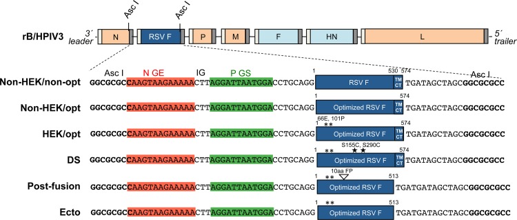
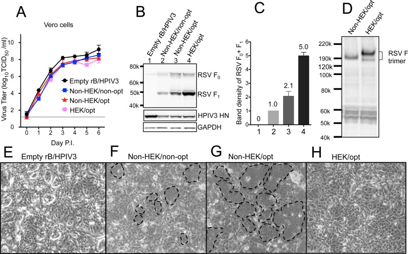
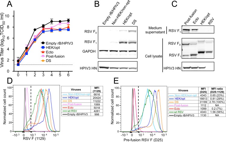
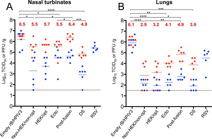
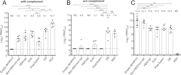
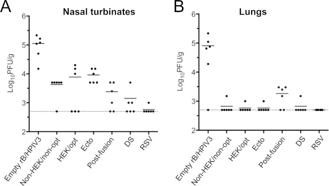
Respiratory syncytial virus (RSV) and human parainfluenza virus type 3 (HPIV3) are the first and second leading viral agents of severe respiratory tract disease in infants and young children worldwide. Vaccines are not available, and an RSV vaccine is particularly needed. A live attenuated chimeric recombinant bovine/human PIV3 (rB/HPIV3) vector expressing the RSV fusion (F) glycoprotein from an added gene has been under development as a bivalent vaccine against RSV and HPIV3. Previous clinical evaluation of this vaccine candidate suggested that increased genetic stability and immunogenicity of the RSV F insert were needed. This was investigated in the present study. RSV F expression was enhanced 5-fold by codon optimization and by modifying the amino acid sequence to be identical to that of an early passage of the original clinical isolate. This conferred a hypofusogenic phenotype that presumably reflects the original isolate. We then compared vectors expressing stabilized prefusion and postfusion versions of RSV F. In a hamster model, prefusion F induced increased quantity and quality of RSV-neutralizing serum antibodies and increased protection against wild-type (wt) RSV challenge. In contrast, a vector expressing the postfusion F was less immunogenic and protective. The genetic stability of the RSV F insert was high and was not affected by enhanced expression or the prefusion or postfusion conformation of RSV F. These studies provide an improved version of the previously well-tolerated rB/HPIV3-RSV F vaccine candidate that induces a superior RSV-neutralizing serum antibody response.

Importance: Respiratory syncytial virus (RSV) and human parainfluenza virus type 3 (HPIV3) are two major causes of pediatric pneumonia and bronchiolitis. The rB/HPIV3 vector expressing RSV F protein is a candidate bivalent live vaccine against HPIV3 and RSV. Previous clinical evaluation indicated the need to increase the immunogenicity and genetic stability of the RSV F insert. Here, we increased RSV F expression by codon optimization and by modifying the RSV F amino acid sequence to conform to that of an early passage of the original isolate. This resulted in a hypofusogenic phenotype, which likely represents the original phenotype before adaptation to cell culture. We also included stabilized versions of prefusion and postfusion RSV F protein. Prefusion RSV F induced a larger quantity and higher quality of RSV-neutralizing serum antibodies and was highly protective. This provides an improved candidate for further clinical evaluation.


Figures

FIG 1
FIG 1
rB/HPIV3 vectors expressing different forms of RSV F. The RSV F ORF (strain A2) was inserted into the rB/HPIV3 vector under the control of a set of added BPIV3 N gene end (GE), intergenic (IG), and P gene start (GS) transcription signals so that RSV F would be expressed as a separate mRNA. With the exception of the top construct (Non-HEK/non-opt), each RSV F insert was codon optimized for human expression (GeneArt; Life Technologies). HEK assignments (66E and 101P) are marked by asterisks. The DS mutations (S155C and S290C) are marked with stars. The Ecto form consists of the RSV F ectodomain (amino acids 1 to 513) lacking the transmembrane and cytoplasmic tail domains. The postfusion form is the Ecto form with a further deletion of the first 10 amino acids of the FP. The viruses were recovered in hamster BSR T7/5 cells as described previously (23) and passaged in rhesus monkey LLC-MK2 cells.
FIG 2
FIG 2
Effects of codon optimization and HEK assignments. (A) Multicycle growth kinetics of rB/HPIV3 vectors in African green monkey Vero cells at 32°C. Triplicate Vero cell monolayers were infected with the indicated rB/HPIV3 vectors at a multiplicity of infection (17) of 0.01 TCID50/cell. Samples were collected at 24-h intervals. Virus titers were determined by serial dilution in LLC-MK2 cells (23) and are expressed as means with standard errors of the mean (SEM) (40). (B) Western blot analysis of RSV F expression in Vero cells, with vector HN protein and cellular GAPDH protein analyzed as controls. Vero cells were infected at an MOI of 10 TCID50/cell at 32°C with the indicated rB/HPIV3 vectors. Total lysates were harvested at 48 h p.i. and subjected to gel electrophoresis under reducing and denaturing conditions, followed by Western blotting, as described previously (23). RSV F0 (70 kDa) is the primary translation product of the F ORF, and RSV F1 (47 kDa) is the larger subunit created when RSV F0 is activated by cleavage. (C) Quantification of RSV F expression. The RSV F1 and F0 band densities (from panel B) were quantified and normalized to that of the Non-HEK/non-opt sample shown at a density value of 1. The means of four independent experiments are shown (numbers above the bars) with the SEM. The numbers on the x axis represent the lanes in Fig. 2B. (D) Mobility of RSV F trimers in polyacrylamide gel electrophoresis under nonreducing conditions. RSV F trimers were detected by Western blotting with rabbit polyclonal antibodies generated by immunizing rabbits with sucrose-purified wt RSV particles. (E to H) Formation of syncytia on Vero cell monolayers infected at an MOI of 10 TCID50/cell with the indicated rB/HPIV3 vectors. The cells were incubated at 32°C for 48 h and photographed using a light microscope at a magnification of ×10. Representative syncytia are marked with dashed lines in panels F and G.
FIG 3
FIG 3
Expression of prefusion DS, postfusion, and Ecto forms of RSV F. (A) Multicycle replication kinetics of the indicated rB/HPIV3 vectors in Vero cells at 32°C. Titration was carried out as described for Fig. 2A. (B) Expression of the prefusion DS form of RSV F. Vero cells were infected with the indicated rB/HPIV3 vectors at an MOI of 10 TCID50/cell and incubated at 37°C for 48 h. Western blot analysis was performed as described for Fig. 2B. (C) Expression and secretion of postfusion, Ecto, and HEK/opt forms of RSV F. Vero cell monolayers were infected with the indicated vectors at an MOI of 10 TCID50/cell or with wt RSV (A2) at an MOI of 10 PFU/cell and incubated at 32°C for 48 h. Western blot analysis was carried out as described for Fig. 2B. (D and E) Cell surface expression of the total and prefusion RSV F protein. Vero cells were infected with the indicated vectors at an MOI of 5 TCID50/cell or with wt RSV at an MOI of 5 PFU/cell and incubated at 32°C. At 48 h p.i., the unpermeabilized cells were stained with RSV F antibody 1129 (D), which reacts with both prefusion and postfusion F, or antibody D25 (E), which is specific for prefusion F. The x axis shows the intensity of RSV F expression, and the y axis is the percentage of the cell count normalized to the maximum count (100%) in a distribution, with the MFI values shown on the right of each histogram. Gating of live RSV F-positive cells used for analysis is indicated with a dashed line.
FIG 4
FIG 4
Replication of rB/HPIV3-RSV-F vectors in hamsters. Golden Syrian hamsters were infected i.n. with 105 TCID50 of the indicated rB/HPIV3 vectors or 106 PFU of wt RSV (A2) in a 0.1-ml inoculum. Hamsters were euthanized (n = 6 per virus per day) on days 3 and 5 postinfection, the nasal turbinates (A) and lungs (B) were collected and homogenized, and the viral titers were determined by limiting dilution on LLC-MK2 (rB/HPIV3 vectors) or Vero (RSV) cells at 32°C. The blue and red dots indicate the titers for individual animals euthanized on days 3 and 5, respectively, and the mean group titer is indicated by a blue or red horizontal line for days 3 and 5, respectively. The mean values of day 5 titers are also shown as red numbers. The limit of detection (LOD) is 1.5 log10 TCID50/g of tissue, indicated with a dotted line. The statistical significance of differences among peak titers on day 5 was determined by one-way analysis of variance (ANOVA) with a Tukey-Kramer test and is indicated by asterisks: *, P ≤ 0.05; **, P ≤ 0.01; ***, P ≤ 0.001; ****, P ≤ 0.0001.
FIG 5
FIG 5
RSV- and HPIV3-neutralizing serum antibody titers induced by rB/HPIV3-RSV F vectors. Hamsters (n = 6 per vector) were infected as described for Fig. 4, and serum samples were collected at 28 days postimmunization. (A and B) RSV-neutralizing antibody titers were determined by a PRNT60 performed on Vero cells at 37°C with (A) and without (B) added guinea pig complement. (C) HPIV3-neutralizing antibody titers were determined by a PRNT60 performed on LLC-MK2 cells at 32°C with added guinea pig complement (23). The height of each bar represents the mean titer, which is shown above the bar; the SEM is indicated by the error bars, and the values for individual animals are shown as dots. The statistical significance of differences among groups was determined as described for Fig. 4; *, P ≤ 0.05; **, P ≤ 0.01; ***, P ≤ 0.001; ****, P ≤ 0.0001; ns, P > 0.05. The detection limit is indicated with a dotted line. ND, the neutralization titer was below the detection limit.
FIG 6
FIG 6
Protection of immunized hamsters against wt RSV challenge. The hamsters (n = 6 per vector) that had been immunized as shown in Fig. 5 were challenged i.n. on day 30 postimmunization with 106 PFU of wt RSV (A2) in a 0.1-ml inoculum. On day 3 postchallenge, the hamsters were euthanized, and nasal turbinates (A) and lungs (B) were collected. The RSV titers in tissue homogenates were determined by plaque assay on Vero cells at 37°C. Each symbol represents the RSV titer for an individual animal, and the mean viral titers of the groups are shown as horizontal lines. The detection limit of the assay was 2.7 log10 PFU/g of tissue, indicated by a dotted line.
FIG 7
FIG 7
Example of fluorescence double-staining plaque titration of a hamster nasal turbinate homogenate. Vero cells were inoculated with a 10-fold serially diluted homogenate of nasal turbinates of hamster number 428 (Table 1), which had been infected with rB/HPIV3 expressing the Ecto form of RSV F. The cells were incubated under a methylcellulose overlay and subjected to double staining for RSV F and HPIV3 antigens. The dilution factors are shown on the left. Shown are staining of duplicate wells for HPIV3 antigens (green) (A) and RSV F (red) (B) (40) and a merged image (plaques expressing RSV F are yellow; plaques that express only HPIV3 antigens are green) (C). The arrows point to a plaque that did not express RSV F in a lower-dilution well. In panel C, there were no green plaques at the dilutions for which individual plaques could be counted (102, 103, and 104), and hence, this sample was scored “100%” for RSV F expression (Table 1), even though sporadic green plaques (e.g., the arrow in panel C) were detected against the yellow background in the 101 dilution.
FIG 8
FIG 8
Example of fluorescence double staining of plaques from a hamster nasal turbinate homogenate with only 14% of recovered vaccine viruses expressing RSV F. Homogenates of nasal turbinates from hamster number 511 (Table 1), which had been infected with rB/HPIV3 expressing the Ecto form of RSV F, were prepared as a 10-fold dilution series, inoculated onto LLC-MK2 cells, incubated under a methylcellulose overlay, and subjected to double staining for RSV F and HPIV3 antigens as described for Fig. 7. The dilution factors are shown on the left. Shown are staining of duplicate wells for HPIV3 antigens (green) (A) and RSV F (red) (40) (B) and a merged image (plaques expressing RSV F are yellow; plaques that express only HPIV3 antigens are green) (C).

References

    1. Nair H, Nokes DJ, Gessner BD, Dherani M, Madhi SA, Singleton RJ, O'Brien KL, Roca A, Wright PF, Bruce N, Chandran A, Theodoratou E, Sutanto A, Sedyaningsih ER, Ngama M, Munywoki PK, Kartasasmita C, Simoes EA, Rudan I, Weber MW, Campbell H. 2010. Global burden of acute lower respiratory infections due to respiratory syncytial virus in young children: a systematic review and meta-analysis. Lancet 375:1545–1555. doi:10.1016/S0140-6736(10)60206-1. - DOI - PMC - PubMed
    1. Ottolini MG, Porter DD, Hemming VG, Prince GA. 2000. Enhanced pulmonary pathology in cotton rats upon challenge after immunization with inactivated parainfluenza virus 3 vaccines. Viral Immunol 13:231–236. doi:10.1089/vim.2000.13.231. - DOI - PubMed
    1. Kim HW, Canchola JG, Brandt CD, Pyles G, Chanock RM, Jensen K, Parrott RH. 1969. Respiratory syncytial virus disease in infants despite prior administration of antigenic inactivated vaccine. Am J Epidemiol 89:422–434. - PubMed
    1. Connors M, Collins PL, Firestone CY, Sotnikov AV, Waitze A, Davis AR, Hung PP, Chanock RM, Murphy BR. 1992. Cotton rats previously immunized with a chimeric RSV FG glycoprotein develop enhanced pulmonary pathology when infected with RSV, a phenomenon not encountered following immunization with vaccinia-RSV recombinants or RSV. Vaccine 10:475–484. doi:10.1016/0264-410X(92)90397-3. - DOI - PubMed
    1. Belshe RB, Newman FK, Tsai TF, Karron RA, Reisinger K, Roberton D, Marshall H, Schwartz R, King J, Henderson FW, Rodriguez W, Severs JM, Wright PF, Keyserling H, Weinberg GA, Bromberg K, Loh R, Sly P, McIntyre P, Ziegler JB, Hackell J, Deatly A, Georgiu A, Paschalis M, Wu SL, Tatem JM, Murphy B, Anderson E. 2004. Phase 2 evaluation of parainfluenza type 3 cold passage mutant 45 live attenuated vaccine in healthy children 6–18 months old. J Infect Dis 189:462–470. doi:10.1086/381184. - DOI - PubMed

Publication types

MeSH terms