

Anatomical Location of the Mesencephalic Locomotor Region and Its Possible Role in Locomotion, Posture, Cataplexy, and Parkinsonism
- PMID: 26157418
- PMCID: PMC4478394
- DOI: 10.3389/fneur.2015.00140
Anatomical Location of the Mesencephalic Locomotor Region and Its Possible Role in Locomotion, Posture, Cataplexy, and Parkinsonism
Abstract
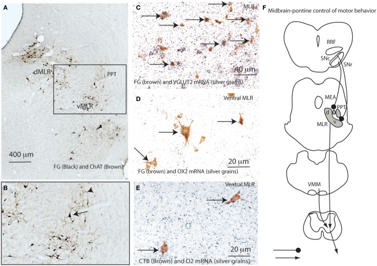
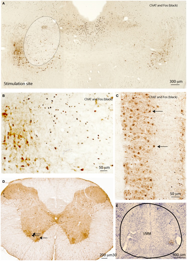
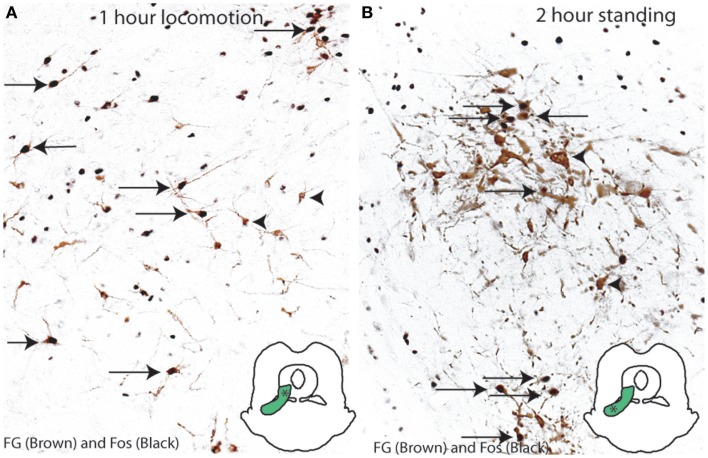
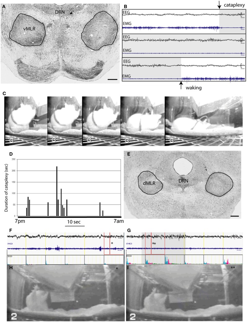
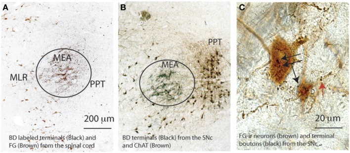
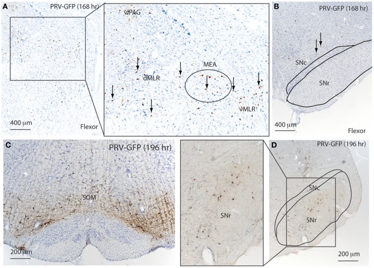
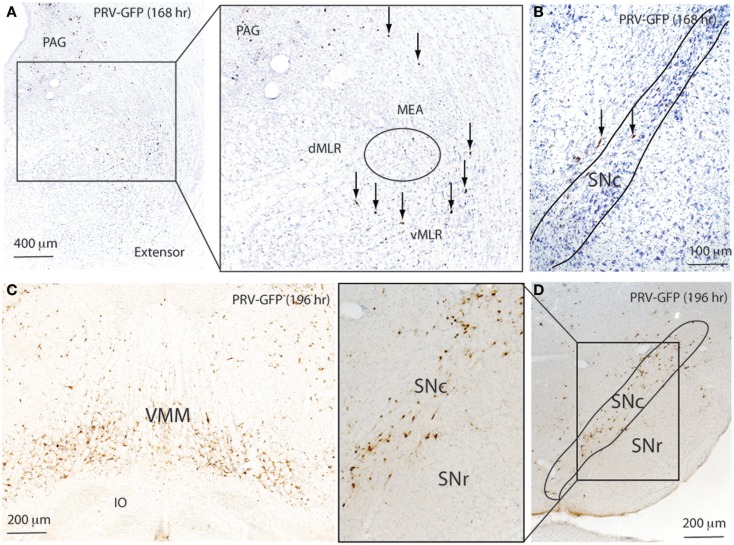
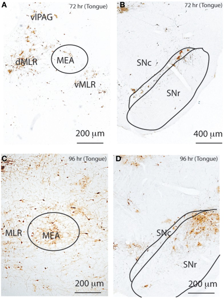
The mesencephalic (or midbrain) locomotor region (MLR) was first described in 1966 by Shik and colleagues, who demonstrated that electrical stimulation of this region induced locomotion in decerebrate (intercollicular transection) cats. The pedunculopontine tegmental nucleus (PPT) cholinergic neurons and midbrain extrapyramidal area (MEA) have been suggested to form the neuroanatomical basis for the MLR, but direct evidence for the role of these structures in locomotor behavior has been lacking. Here, we tested the hypothesis that the MLR is composed of non-cholinergic spinally projecting cells in the lateral pontine tegmentum. Our results showed that putative MLR neurons medial to the PPT and MEA in rats were non-cholinergic, glutamatergic, and express the orexin (hypocretin) type 2 receptors. Fos mapping correlated with motor behaviors revealed that the dorsal and ventral MLR are activated, respectively, in association with locomotion and an erect posture. Consistent with these findings, chemical stimulation of the dorsal MLR produced locomotion, whereas stimulation of the ventral MLR caused standing. Lesions of the MLR (dorsal and ventral regions together) resulted in cataplexy and episodic immobility of gait. Finally, trans-neuronal tracing with pseudorabies virus demonstrated disynaptic input to the MLR from the substantia nigra via the MEA. These findings offer a new perspective on the neuroanatomic basis of the MLR, and suggest that MLR dysfunction may contribute to the postural and gait abnormalities in Parkinsonism.
Keywords: basal ganglia; cataplexy; dopamine; pontine motor; sleep.
Figures
Similar articles
-
Movement- and behavioral state-dependent activity of pontine reticulospinal neurons.Neuroscience. 2012 Sep 27;221:125-39. doi: 10.1016/j.neuroscience.2012.06.069. Epub 2012 Jul 13. Neuroscience. 2012. PMID: 22796072 Free PMC article.
-
Orexinergic projections to the cat midbrain mediate alternation of emotional behavioural states from locomotion to cataplexy.J Physiol. 2005 Nov 1;568(Pt 3):1003-20. doi: 10.1113/jphysiol.2005.085829. Epub 2005 Aug 25. J Physiol. 2005. PMID: 16123113 Free PMC article.
-
Activation of Brainstem Neurons During Mesencephalic Locomotor Region-Evoked Locomotion in the Cat.Front Syst Neurosci. 2019 Nov 14;13:69. doi: 10.3389/fnsys.2019.00069. eCollection 2019. Front Syst Neurosci. 2019. PMID: 31798423 Free PMC article.
-
Brainstem control of locomotion and muscle tone with special reference to the role of the mesopontine tegmentum and medullary reticulospinal systems.J Neural Transm (Vienna). 2016 Jul;123(7):695-729. doi: 10.1007/s00702-015-1475-4. Epub 2015 Oct 26. J Neural Transm (Vienna). 2016. PMID: 26497023 Free PMC article. Review.
-
Initiation of locomotion in lampreys.Brain Res Rev. 2008 Jan;57(1):172-82. doi: 10.1016/j.brainresrev.2007.07.016. Epub 2007 Aug 22. Brain Res Rev. 2008. PMID: 17916380 Review.
Cited by
-
The Relationship between Saccades and Locomotion.J Mov Disord. 2018 Sep;11(3):93-106. doi: 10.14802/jmd.18018. Epub 2018 Aug 9. J Mov Disord. 2018. PMID: 30086615 Free PMC article.
-
Differential spatiotemporal gait effects with frequency and dopaminergic modulation in STN-DBS.Front Aging Neurosci. 2023 Sep 29;15:1206533. doi: 10.3389/fnagi.2023.1206533. eCollection 2023. Front Aging Neurosci. 2023. PMID: 37842127 Free PMC article.
-
Stereological estimations and neurochemical characterization of neurons expressing GABAA and GABAB receptors in the rat pedunculopontine and laterodorsal tegmental nuclei.Brain Struct Funct. 2022 Jan;227(1):89-110. doi: 10.1007/s00429-021-02375-9. Epub 2021 Sep 12. Brain Struct Funct. 2022. PMID: 34510281 Free PMC article.
-
Freezing of Gait in Parkinson's Disease: Invasive and Noninvasive Neuromodulation.Neuromodulation. 2021 Jul;24(5):829-842. doi: 10.1111/ner.13347. Epub 2020 Dec 26. Neuromodulation. 2021. PMID: 33368872 Free PMC article. Review.
-
Orexinergic Modulation of Spinal Motor Activity in the Neonatal Mouse Spinal Cord.eNeuro. 2018 Nov 8;5(5):ENEURO.0226-18.2018. doi: 10.1523/ENEURO.0226-18.2018. eCollection 2018 Sep-Oct. eNeuro. 2018. PMID: 30417080 Free PMC article.
References
-
- Sprague JM, Chambers WW. Control of posture by reticular formation and cerebellum in the intract, anesthetized and unanesthetized and in the decerebrated cat. Am J Physiol (1954) 176:52–64. - PubMed
-
- Shik ML, Severin FV, Orlovskii GN. [Control of walking and running by means of electric stimulation of the midbrain]. Biofizika (1966) 11:659–66. - PubMed
-
- Berman AL. The Brain Stem of the Cat; A Cytoarchitectonic Atlas with Stereotaxic Coordinates. Madison: University of Wisconsin Press; (1968).
Grants and funding
LinkOut - more resources
Full Text Sources
Other Literature Sources
Miscellaneous