

PE11 (Rv1169c) selectively alters fatty acid components of Mycobacterium smegmatis and host cell interleukin-6 level accompanied with cell death
- PMID: 26157429
- PMCID: PMC4477156
- DOI: 10.3389/fmicb.2015.00613
PE11 (Rv1169c) selectively alters fatty acid components of Mycobacterium smegmatis and host cell interleukin-6 level accompanied with cell death
Abstract
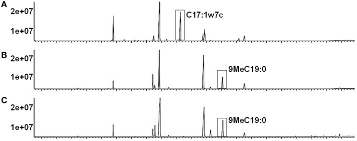
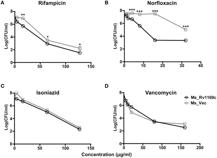
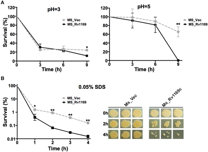
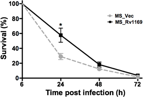
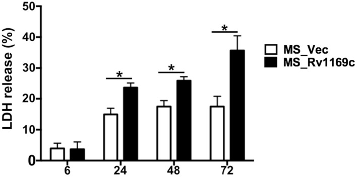
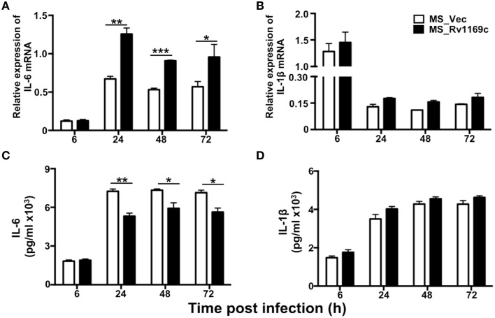
PE/PPE family proteins, named after their conserved PE (Pro-Glu) and PPE (Pro-Pro-Glu) domains of N-terminal, are most intriguing aspects of pathologic mycobacterial genome. The roles of most members of this family remain unknown, although selected genes of this family are related to the virulence of Mycobacterium tuberculosis. In order to decipher the role of Rv1169c, the Mycobacterium smegmatis strain heterologous expressed this ORF was constructed and identified that Rv1169c was a cell wall associated protein with a novel function in modifying the cell wall fatty acids. The growth of Rv1169c expressing strain was affected under surface stress, acidic condition and antibiotics treatment. M. smegmatis expressing Rv1169c induced necrotic cell death of macrophage after infection and significantly decreased interlukin-6 production compared to controls. In general, these results underscore a proposing role of Rv1169c in virulence of M. tuberculosis, as it's role in the susceptibility of anti-mycobacteria factors caused by modified cell wall fatty acid, and the induced necrotic cell death by Rv1169c is crucial for M. tuberculosis virulence during infection.
Keywords: Mycobacterium tuberculosis; PE family; Rv1169c; cell death; cell wall integrality; fatty acid components; interleukin-6.
Figures
Similar articles
-
PE11, a PE/PPE family protein of Mycobacterium tuberculosis is involved in cell wall remodeling and virulence.Sci Rep. 2016 Feb 23;6:21624. doi: 10.1038/srep21624. Sci Rep. 2016. PMID: 26902658 Free PMC article.
-
Down-regulation of PE11, a cell wall associated esterase, enhances the biofilm growth of Mycobacterium tuberculosis and reduces cell wall virulence lipid levels.Microbiology (Reading). 2017 Jan;163(1):52-61. doi: 10.1099/mic.0.000417. Microbiology (Reading). 2017. PMID: 28198348
-
Mycobacterium tuberculosis PE13 (Rv1195) manipulates the host cell fate via p38-ERK-NF-κB axis and apoptosis.Apoptosis. 2016 Jul;21(7):795-808. doi: 10.1007/s10495-016-1249-y. Apoptosis. 2016. PMID: 27147522
-
Expression and regulatory networks of Mycobacterium tuberculosis PE/PPE family antigens.J Cell Physiol. 2019 Jun;234(6):7742-7751. doi: 10.1002/jcp.27608. Epub 2018 Nov 27. J Cell Physiol. 2019. PMID: 30478834 Review.
-
Phylogeny to function: PE/PPE protein evolution and impact on Mycobacterium tuberculosis pathogenicity.Mol Microbiol. 2015 Jun;96(5):901-16. doi: 10.1111/mmi.12981. Epub 2015 Mar 30. Mol Microbiol. 2015. PMID: 25727695 Review.
Cited by
-
The Mycobacterium tuberculosis PE_PGRS Protein Family Acts as an Immunological Decoy to Subvert Host Immune Response.Int J Mol Sci. 2022 Jan 4;23(1):525. doi: 10.3390/ijms23010525. Int J Mol Sci. 2022. PMID: 35008950 Free PMC article. Review.
-
Expression and purification of a gene encoding a 9.7 kDa PE protein of Mycobacterium avium subsp. paratuberculosis.3 Biotech. 2016 Dec;6(2):198. doi: 10.1007/s13205-016-0506-7. Epub 2016 Sep 15. 3 Biotech. 2016. PMID: 28330270 Free PMC article.
-
Mycobacterium PPE31 Contributes to Host Cell Death.Front Cell Infect Microbiol. 2021 Apr 13;11:629836. doi: 10.3389/fcimb.2021.629836. eCollection 2021. Front Cell Infect Microbiol. 2021. PMID: 33928042 Free PMC article.
-
Novel role of PE_PGRS47 in the alteration of mycobacterial cell wall integrity and drug resistance.Arch Microbiol. 2023 Apr 6;205(5):174. doi: 10.1007/s00203-023-03515-x. Arch Microbiol. 2023. PMID: 37022460
-
Modulation of Trehalose Dimycolate and Immune System by Rv0774c Protein Enhanced the Intracellular Survival of Mycobacterium smegmatis in Human Macrophages Cell Line.Front Cell Infect Microbiol. 2017 Jun 30;7:289. doi: 10.3389/fcimb.2017.00289. eCollection 2017. Front Cell Infect Microbiol. 2017. PMID: 28713776 Free PMC article.
References
-
- Bai X., Kinney W. H., Su W. L., Bai A., Ovrutsky A. R., Honda J. R., et al. . (2015). Caspase-3-independent apoptotic pathways contribute to interleukin-32gamma-mediated control of Mycobacterium tuberculosis infection in THP-1 cells. BMC Microbiol. 15:39. 10.1186/s12866-015-0366-z - DOI - PMC - PubMed
-
- Bansal K., Sinha A. Y., Ghorpade D. S., Togarsimalemath S. K., Patil S. A., Kaveri S. V., et al. . (2010). Src homology 3-interacting domain of Rv1917c of Mycobacterium tuberculosis induces selective maturation of human dendritic cells by regulating PI3K-MAPK-NF-kappaB signaling and drives Th2 immune responses. J. Biol. Chem. 285, 36511–36522. 10.1074/jbc.M110.158055 - DOI - PMC - PubMed
LinkOut - more resources
Full Text Sources
Other Literature Sources
Molecular Biology Databases