. 2015 Jun 23:6:641.
doi: 10.3389/fmicb.2015.00641. eCollection 2015.

High-dose rifampicin kills persisters, shortens treatment duration, and reduces relapse rate in vitro and in vivo

Affiliations

High-dose rifampicin kills persisters, shortens treatment duration, and reduces relapse rate in vitro and in vivo

Yanmin Hu et al. Front Microbiol. .

Abstract

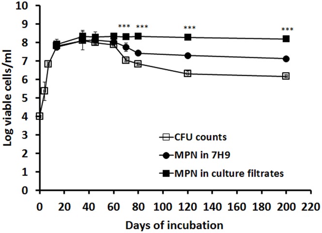
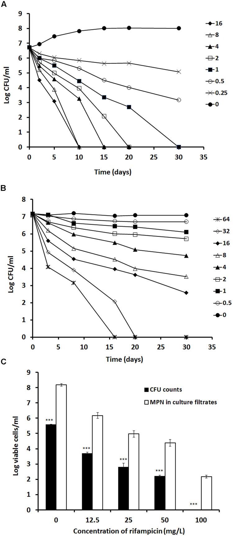
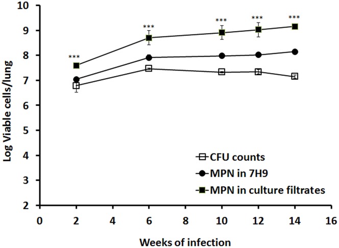
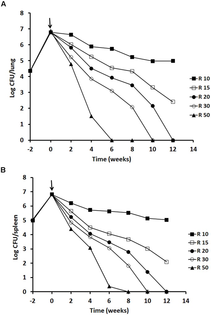
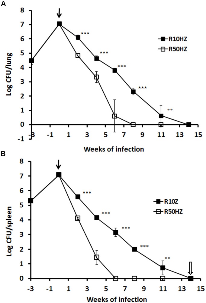
Although high-dose rifampicin holds promise for improving tuberculosis control by potentially shortening treatment duration, these effects attributed to eradication of persistent bacteria are unclear. The presence of persistent Mycobacterium tuberculosis was examined using resuscitation promoting factors (RPFs) in both in vitro hypoxia and in vivo murine tuberculosis models before and after treatment with incremental doses of rifampicin. Pharmacokinetic parameters and dose-dependent profile of rifampicin in the murine model were determined. The Cornell mouse model was used to test efficacy of high-dose rifampicin in combination with isoniazid and pyrazinamide and to measure relapse rate. There were large numbers of RPF-dependent persisters in vitro and in vivo. Stationary phase cultures were tolerant to rifampicin while higher concentrations of rifampicin eradicated plate count positive but not RPF-dependent persistent bacteria. In murine infection model, incremental doses of rifampicin exhibited a dose-dependent eradication of RPF-dependent persisters. Increasing the dose of rifampicin significantly reduced the risk of antibiotic resistance emergence. In Cornell model, mice treated with high-dose rifampicin regimen resulted in faster visceral clearance; organs were M. tuberculosis free 8 weeks post-treatment compared to 14 weeks with standard-dose rifampicin regimen. Organ sterility, plate count and RPF-dependent persister negative, was achieved. There was no disease relapse compared to the standard dose regimen (87.5%). High-dose rifampicin therapy results in eradication of RPF-dependent persisters, allowing shorter treatment duration without disease relapse. Optimizing rifampicin to its maximal efficacy with acceptable side-effect profiles will provide valuable information in human studies and can potentially improve current tuberculosis chemotherapy.

Keywords: Mycobacterium tuberculosis; mouse model; persistence; resuscitation promoting factors; rifampicin.


Figures

FIGURE 1
FIGURE 1
Resuscitation of RPF-dependent tubercle bacilli from the in vitro hypoxic model of M. tuberculosis. M. tuberculosis H37Rv was grown in 7H9 medium without disturbance for 200 days. CFU counts were performed at different time points in triplicate (line with empty squares). MPN counts from cultures of 14, 35, 45, 60, 70, 80, 120, and 200 days were performed with 7H9 medium (line with solid circles) or the culture filtrate (line with solid squares). These experiments were performed three times with reproducible results. ANOVA analysis demonstrated that there were significant differences amongst CFU counts, 7H9 broth counts and RPF broth counts (∗∗∗P < 0.001 to 0.0001).
FIGURE 2
FIGURE 2
Determination of rifampicin activity against log-phase and stationary-phase M. tuberculosis using time kill curve analysis. Rifampicin at the concentration from 16 to 0 mg/L were incubated with a 7 days log-phase culture (A) and rifampicin at the concentration from 64 to 0 mg/L were incubated with the 60 days stationary-phase culture (B). CFU counts were estimated at different time points. A 100-days culture was treated with 12.5, 25, 50, and 100 mg/L of rifampicin for 5 days. After removal of rifampicin, the treated culture were subject to CFU counting on agar plates and MPN counting with the culture filtrates (C). These experiments were performed three times with reproducible results. Statistical analysis demonstrated that the decline of CFU counts after treatment with different concentrations of rifampicin was significant, P < 0.0008 (ANOVA), for the log-phase culture (A), P < 0.001 (ANOVA) for the stationary phase culture (B) and P < 0.001 (∗∗∗ Student’s t-test) for the 100-days culture (C).
FIGURE 3
FIGURE 3
Resuscitation of M. tuberculosis grown in mouse lungs. BALB/c mice were infected with M. tuberculosis. Viability of the bacilli in lung was determined by CFU counting and MPN counting with 7H9 or the culture filtrates at 2, 6, 10, 12, and 14 weeks post-infection. The results have been performed twice with reproducible results. ANOVA analysis demonstrated that there were significant differences amongst CFU counts, 7H9 broth counts and RPF broth counts (∗∗∗P < 0.001) in mice.
FIGURE 4
FIGURE 4
Viability of M. tuberculosis H37Rv in BALB/c mice after rifampicin treatment. The results of a single experiment are shown with viability expressed as log CFU counts per lung (A) and spleen (B). Mice were infected intravenously and the infection was allowed to progress for 2 weeks prior to treatment with rifampicin at 10, 15, 20, 30 and 50 mg/kg indicated as a solid arrow for 12 weeks (time weeks 0–12). At week 2, 4, 6, 8, and 12 of post-treatment, four mice from each group were sacrificed for CFU counting. Data of the mice containing rifampicin-resistant strains for the 10 mg/kg group was excluded. ANOVA analysis demonstrated that the decline of CFU counts after treatment with different concentrations of rifampicin was significant in lungs P < 0.02 and in spleens P < 0.04.
FIGURE 5
FIGURE 5
Viability of M. tuberculosis H37Rv in the mouse Cornell model of dormancy. The results of a single experiment are shown with viability expressed as log CFU counts per lung (A) and spleen (B). Mice were infected intravenously at week –3 and the infection was allowed to progress for 3 weeks prior to treatment with isoniazid and pyrazinamide with rifampicin added at 10 or 50 mg/kg indicated as a solid arrow for 14 weeks (time weeks 0–14). At week 2, 4, 6, 8, 11, and 14 of post-treatment, CFU counts in the organs from each group were estimated. Steroid treatment was started immediately after the termination of 14 weeks of antibiotic treatment as indicated with an empty arrow. The experiment has been performed twice with reproducible results. Statistical analysis demonstrated that the decline of CFU counts in lungs and spleens was significant between low and high-dose rifampicin regiments, ∗∗∗P < 0.0001 or ∗∗P < 0.02 (Student’s t-test).

Similar articles

Cited by

References

    1. Abdool Karim S. S., Naidoo K., Grobler A., Padayatchi N., Baxter C., Gray A., et al. (2010). Timing of initiation of antiretroviral drugs during tuberculosis therapy. N. Engl. J. Med. 362 697–706. 10.1056/NEJMoa0905848 - DOI - PMC - PubMed
    1. Boeree M. J., Diacon A., Dawson R., Van Balen G. P., Venter A., Du Bois J., et al. (2013). What is the Right Dose of Rifampin? A Dose Escalating Study. Available at: http://www.atsjournals.org/doi/abs/10.1164/ajrccm-conference.2013.187.1_... - DOI
    1. Calver A. D., Falmer A. A., Murray M., Strauss O. J., Streicher E. M., Hanekom M., et al. (2010). Emergence of increased resistance and extensively drug-resistant tuberculosis despite treatment adherence, South Africa. Emerg. Infect. Dis. 16 264–271. 10.3201/eid1602.090968 - DOI - PMC - PubMed
    1. Coates A. R., Hu Y., Jindani A., Mitchison D. A. (2013). Contradictory results with high-dosage rifamycin in mice and humans. Antimicrob. Agents Chemother. 57 1103 10.1128/AAC.01705-12 - DOI - PMC - PubMed
    1. de Steenwinkel J. E., Aarnoutse R. E., de Knegt G. J., Ten Kate M. T., Teulen M., Verbrugh H. A., et al. (2013). Optimization of the rifampin dosage to improve the therapeutic efficacy in tuberculosis treatment, using a murine model. Am. J. Respir. Crit. Care Med. 187 1127–1134. 10.1164/rccm.201207-1210OC - DOI - PubMed