

MethPed: a DNA methylation classifier tool for the identification of pediatric brain tumor subtypes
- PMID: 26157508
- PMCID: PMC4495799
- DOI: 10.1186/s13148-015-0103-3
MethPed: a DNA methylation classifier tool for the identification of pediatric brain tumor subtypes
Abstract
Background: Classification of pediatric tumors into biologically defined subtypes is challenging, and multifaceted approaches are needed. For this aim, we developed a diagnostic classifier based on DNA methylation profiles.
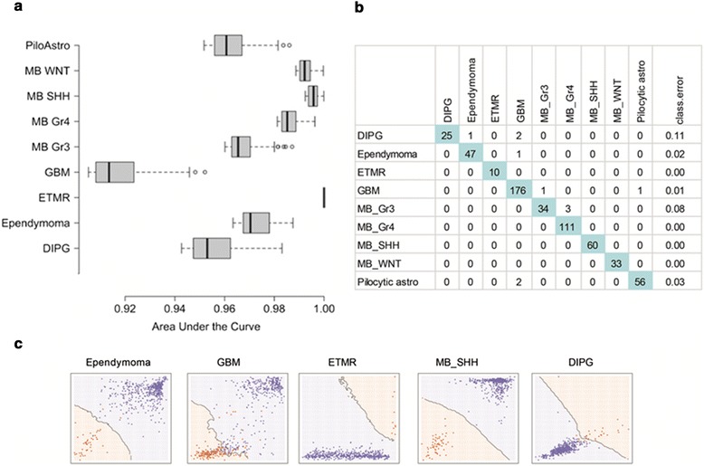
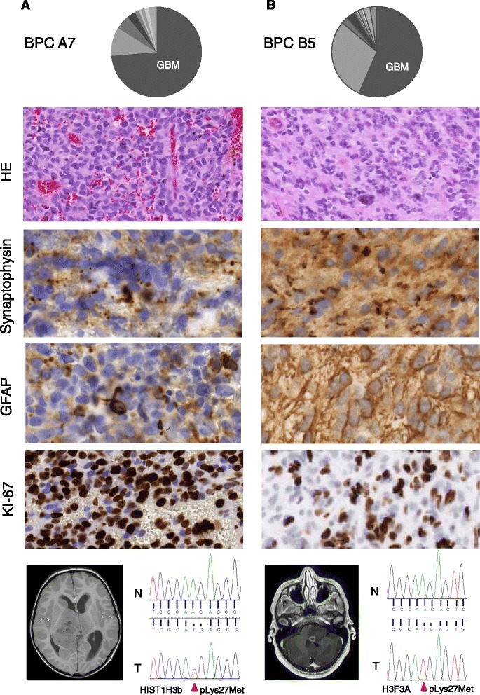
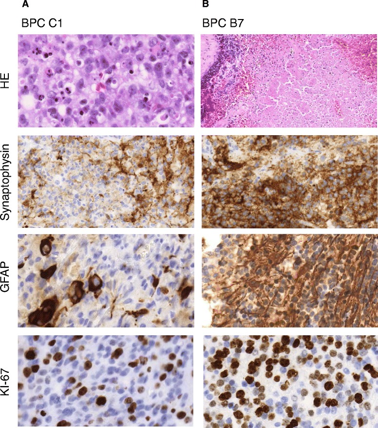
Results: Methylation data generated by the Illumina Infinium HumanMethylation 450 BeadChip arrays were downloaded from the Gene Expression Omnibus (n = 472). Using the data, we built MethPed, which is a multiclass random forest algorithm, based on DNA methylation profiles from nine subgroups of pediatric brain tumors. DNA from 18 regional samples was used to validate MethPed. MethPed was additionally applied to a set of 28 publically available tumors with the heterogeneous diagnosis PNET. MethPed could successfully separate individual histology tumor types at a very high accuracy (κ = 0.98). Analysis of a regional cohort demonstrated the clinical benefit of MethPed, as confirmation of diagnosis of tumors with clear histology but also identified possible differential diagnoses in tumors with complicated and mixed type morphology.
Conclusions: We demonstrate the utility of methylation profiling of pediatric brain tumors and offer MethPed as an easy-to-use toolbox that allows researchers and clinical diagnosticians to test single samples as well as large cohorts for subclass prediction of pediatric brain tumors. This will immediately aid clinical practice and importantly increase our molecular knowledge of these tumors for further therapeutic development.
Keywords: 450 K; Astrocytoma; Classifier (classification tool); DNA methylation; Ependymoma; GBM; Medulloblastoma; MethPed; PNET; Random forest.
Figures
Similar articles
-
MethPed: an R package for the identification of pediatric brain tumor subtypes.BMC Bioinformatics. 2016 Jul 2;17(1):262. doi: 10.1186/s12859-016-1144-0. BMC Bioinformatics. 2016. PMID: 27370569 Free PMC article.
-
Development of a Machine Learning Classifier for Brain Tumors Diagnosis Based on DNA Methylation Profile.Front Bioinform. 2021 Nov 8;1:744345. doi: 10.3389/fbinf.2021.744345. eCollection 2021. Front Bioinform. 2021. PMID: 36303797 Free PMC article.
-
Sarcoma classification by DNA methylation profiling in clinical everyday life: the Charité experience.Clin Epigenetics. 2022 Nov 15;14(1):149. doi: 10.1186/s13148-022-01365-w. Clin Epigenetics. 2022. PMID: 36380356 Free PMC article.
-
Current status of DNA methylation profiling in neuro-oncology as a diagnostic support tool: A review.Neurooncol Pract. 2023 Jul 22;10(6):518-526. doi: 10.1093/nop/npad040. eCollection 2023 Dec. Neurooncol Pract. 2023. PMID: 38009119 Free PMC article. Review.
-
Brain Tumor Classification by Methylation Profile.J Korean Med Sci. 2023 Nov 6;38(43):e356. doi: 10.3346/jkms.2023.38.e356. J Korean Med Sci. 2023. PMID: 37935168 Free PMC article. Review.
Cited by
-
Comparison of DNA methylation based classification models for precision diagnostics of central nervous system tumors.NPJ Precis Oncol. 2024 Oct 2;8(1):218. doi: 10.1038/s41698-024-00718-3. NPJ Precis Oncol. 2024. PMID: 39358389 Free PMC article.
-
DNA methylation profiling improves routine diagnosis of paediatric central nervous system tumours: A prospective population-based study.Neuropathol Appl Neurobiol. 2022 Oct;48(6):e12838. doi: 10.1111/nan.12838. Epub 2022 Aug 3. Neuropathol Appl Neurobiol. 2022. PMID: 35892159 Free PMC article.
-
Batch-normalization of cerebellar and medulloblastoma gene expression datasets utilizing empirically defined negative control genes.Bioinformatics. 2019 Sep 15;35(18):3357-3364. doi: 10.1093/bioinformatics/btz066. Bioinformatics. 2019. PMID: 30715209 Free PMC article.
-
Cell line-based xenograft mouse model of paediatric glioma stem cells mirrors the clinical course of the patient.Carcinogenesis. 2018 Oct 8;39(10):1304-1309. doi: 10.1093/carcin/bgy091. Carcinogenesis. 2018. PMID: 29982329 Free PMC article.
-
Brain tumour diagnostics using a DNA methylation-based classifier as a diagnostic support tool.Neuropathol Appl Neurobiol. 2020 Aug;46(5):478-492. doi: 10.1111/nan.12610. Epub 2020 Apr 7. Neuropathol Appl Neurobiol. 2020. PMID: 32072658 Free PMC article.
References
-
- Sexton-Oates A, MacGregor D, Dodgshun A, Saffery R. The potential for epigenetic analysis of paediatric CNS tumours to improve diagnosis, treatment and prognosis. Ann Oncol. 2015. doi:10.1093/annonc/mdv024. - PubMed
LinkOut - more resources
Full Text Sources
Other Literature Sources
Molecular Biology Databases
