. 2015 Jun 23:2:32.
doi: 10.3389/fmolb.2015.00032. eCollection 2015.

Structural modeling of the N-terminal signal-receiving domain of IκBα

Affiliations

Structural modeling of the N-terminal signal-receiving domain of IκBα

Samira Yazdi et al. Front Mol Biosci. .

Abstract

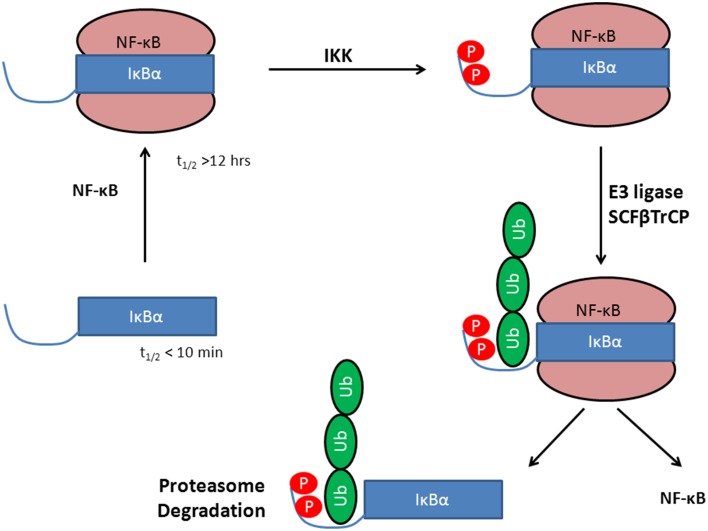
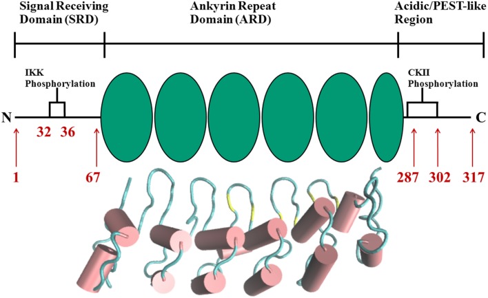
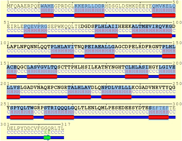
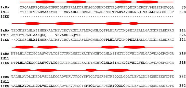
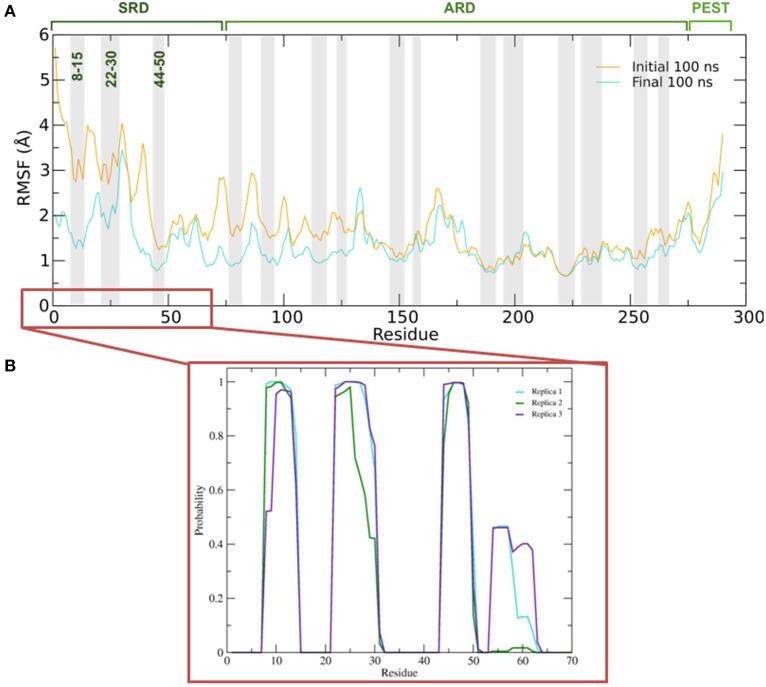
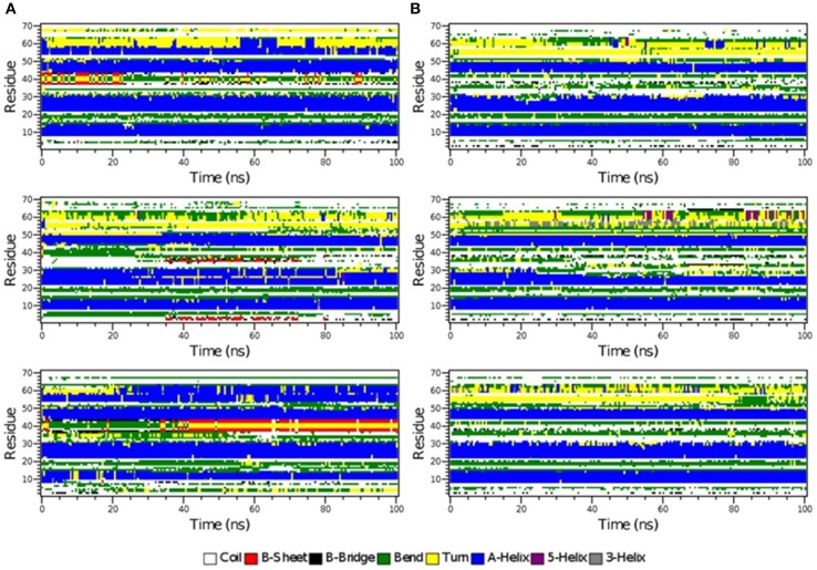
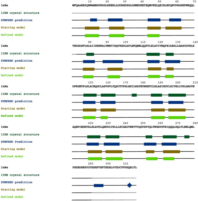
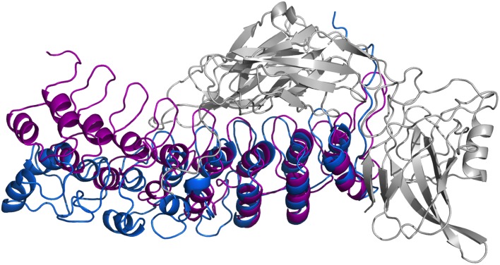
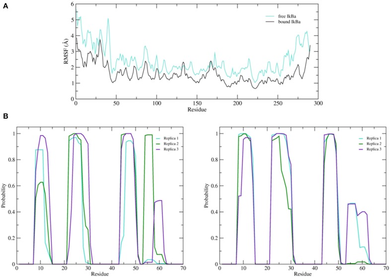
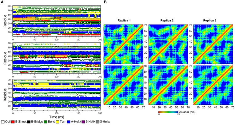
The transcription factor nuclear factor-κB (NF-κB) exerts essential roles in many biological processes including cell growth, apoptosis and innate and adaptive immunity. The NF-κB inhibitor (IκBα) retains NF-κB in the cytoplasm and thus inhibits nuclear localization of NF-κB and its association with DNA. Recent protein crystal structures of the C-terminal part of IκBα in complex with NF-κB provided insights into the protein-protein interactions but could not reveal structural details about the N-terminal signal receiving domain (SRD). The SRD of IκBα contains a degron, formed following phosphorylation by IκB kinases (IKK). In current protein X-ray structures, however, the SRD is not resolved and assumed to be disordered. Here, we combined secondary structure annotation and domain threading followed by long molecular dynamics (MD) simulations and showed that the SRD possesses well-defined secondary structure elements. We show that the SRD contains 3 additional stable α-helices supplementing the six ARDs present in crystallized IκBα. The IκBα/NF-κB protein-protein complex remained intact and stable during the entire simulations. Also in solution, free IκBα retains its structural integrity. Differences in structural topology and dynamics were observed by comparing the structures of NF-κB free and NF-κB bound IκBα-complex. This study paves the way for investigating the signaling properties of the SRD in the IκBα degron. A detailed atomic scale understanding of molecular mechanism of NF-κB activation, regulation and the protein-protein interactions may assist to design and develop novel chronic inflammation modulators.

Keywords: IκBα; N-terminal extension; NF-κB; molecular dynamics simulation; protein-protein complex refinement; secondary structure prediction; signal receiving domain; signal transduction.


Figures

Figure 1
Figure 1
Stabilization of IκBα upon protein-protein complex formation with the transcription factor NF-κB. Phosphorylation by IKK leads to a degron, recognized by SCF(β-TrCP) and subsequent IκBα ubiquitination by E3. This activates NF-κB whereas IκBα is degraded in the 26S proteasome.
Figure 2
Figure 2
Schematic representation of protein domains in IκBα. The N-terminal SRD is the site of phosphorylation by IKK and subsequent ubiquitination by the SCF (β-TrCP) E3 ligase, six ankyrin repeat units make up the central ARD domain. The ankyrin repeat units in the protein crystal structure of IκBα in complex with NF-κB (pdb entry 1IKN) are shown as a cartoon representation. Indicated for the C-terminal PEST-like region are the CKII sites of phosphorylation. The diversity of binding sites, the great variability of κB-sites in the DNA motif and the existence of suppressive and inductive NF-κB dimers lead to a complexity and versatility of the downstream signaling network.
Figure 3
Figure 3
Consensus secondary structure annotation of full length IκBα (residues 1–317). Truncated, as crystallized IκBα from 1IKN (residues 73–293) is shown in bold letters. The six ankyrin repeat units of the ARD are recovered, correctly annotated and positioned. Two additional α-helix-loop-α-helix regions were detected in the N-terminal SRD. The C-terminal PEST domain displays less structural features.
Figure 4
Figure 4
Two-template sequence alignment used for the generation of a composite structural model of the full-sequence IκBα. The bold segments in each template correspond to the α-helical regions forming the ankyrin repeat units present in the crystal structures of 1IKN and 1N11. The curved boxes in red display the helical segments in our generated structural model of IκBα.
Figure 5
Figure 5
(A) Average root mean square fluctuations (RMSF) of the backbone of IκBα for the initial and final 100 ns of the simulation. Shaded areas depict α-helical regions at the end of the three independent 200 ns simulation periods. (B) Probability distribution of α-helix formation of the first 70 residues of the SRD of IκBα in complex with NF-κB.
Figure 6
Figure 6
The secondary structure elements of the first 70 N-terminal residues of IκBα in complex with NF-κB as calculated by DSSP for the three system replicas during the initial 100ns (A) and final 100 ns (B) of the simulation.
Figure 7
Figure 7
A graphical map of the secondary structure elements of IκBα, displayed on its complete sequence. The boxes highlight the α-helical regions, and the arrows indicate β-strands. Dark green designates secondary structures determined in the crystal structure 1IKN, blue denotes secondary structures predicted by SYMPRED, and brown and fluorescent green indicate secondary structures suggested by our initial and refined structural models.
Figure 8
Figure 8
Ribbon diagrams of the three-dimensional initial structure (purple) and the refined structure after a 200 ns MD simulation (blue) of IκBα. The structures are shown in comparison by superpositioning IκBα's binding partner NF-κB (gray).
Figure 9
Figure 9
(A) Average root mean square fluctuations (RMSF) of the backbone of the free IκBα (cyan) in comparison to the one in complex with NF-κB (black). (B) Probability distributions of α-helix formation of the first 70 residues of the SRD. Left: free IκBα. Right: IκBα in complex with NF-κB.
Figure 10
Figure 10
(A) The secondary structure elements of the first 70 N-terminal residues of free IκBα calculated by DSSP for the three system replicas for the entire simulation. (B) Interatomic distance matrices for the first 70 N-terminal residues of free IκBα (top) and in complex with NF-κB (bottom).

References

    1. Bouma G., Strober W. (2003). The immunological and genetic basis of inflammatory bowel disease. Nat. Rev. Immunol. 37, 521–533. 10.1038/nri1132 - DOI - PubMed
    1. Bradley P., Misura K. M. S., Baker D. (2005). Toward high-resolution de novo structure prediction for small proteins. Science 309, 1868–1871. 10.1126/science.1113801 - DOI - PubMed
    1. Cervantes C. F., Markwick P. R. L., Sue S. C., McCammon J. A., Dyson H. J., Komives E. A. (2009). Functional dynamics of the folded ankyrin repeats of I κB α revealed by nuclear magnetic resonance. Biochemistry 48, 8023–8031. 10.1021/bi900712r - DOI - PMC - PubMed
    1. Chen F. E., Huang D. B., Chen Y. Q., Ghosh G. (1998). Crystal structure of p50/p65 heterodimer of transcription factor NF-κB bound to DNA. Nature 391, 410–413. 10.1038/34356 - DOI - PubMed
    1. Cheng J., Randall A. Z., Sweredoski M. J., Baldi P. (2005). SCRATCH: a protein structure and structural feature prediction server. Nucleic Acids Res. 33, W72–W76. 10.1093/nar/gki396 - DOI - PMC - PubMed

LinkOut - more resources