. 2015 Apr;2(2):021013.
doi: 10.1117/1.NPh.2.2.021013. Epub 2015 Mar 31.

All-optical mapping of barrel cortex circuits based on simultaneous voltage-sensitive dye imaging and channelrhodopsin-mediated photostimulation

Affiliations

All-optical mapping of barrel cortex circuits based on simultaneous voltage-sensitive dye imaging and channelrhodopsin-mediated photostimulation

Shun Qiang Lo et al. Neurophotonics. 2015 Apr.

Abstract

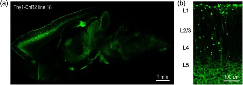
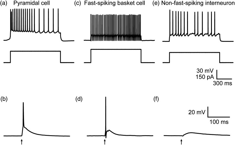
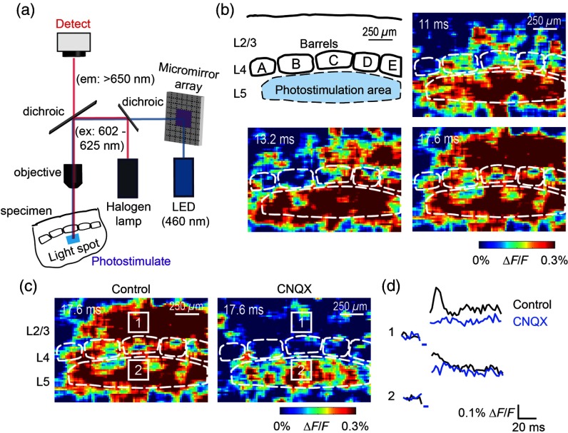
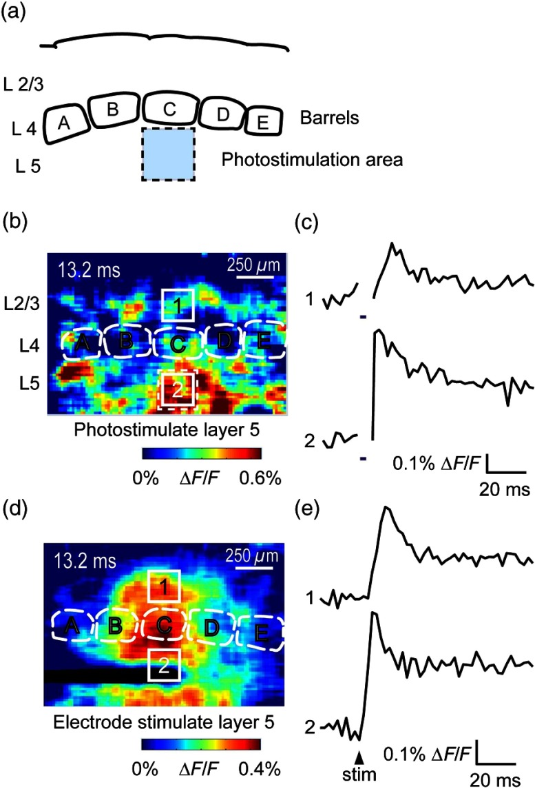
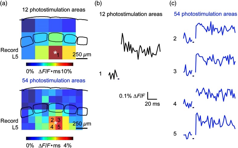
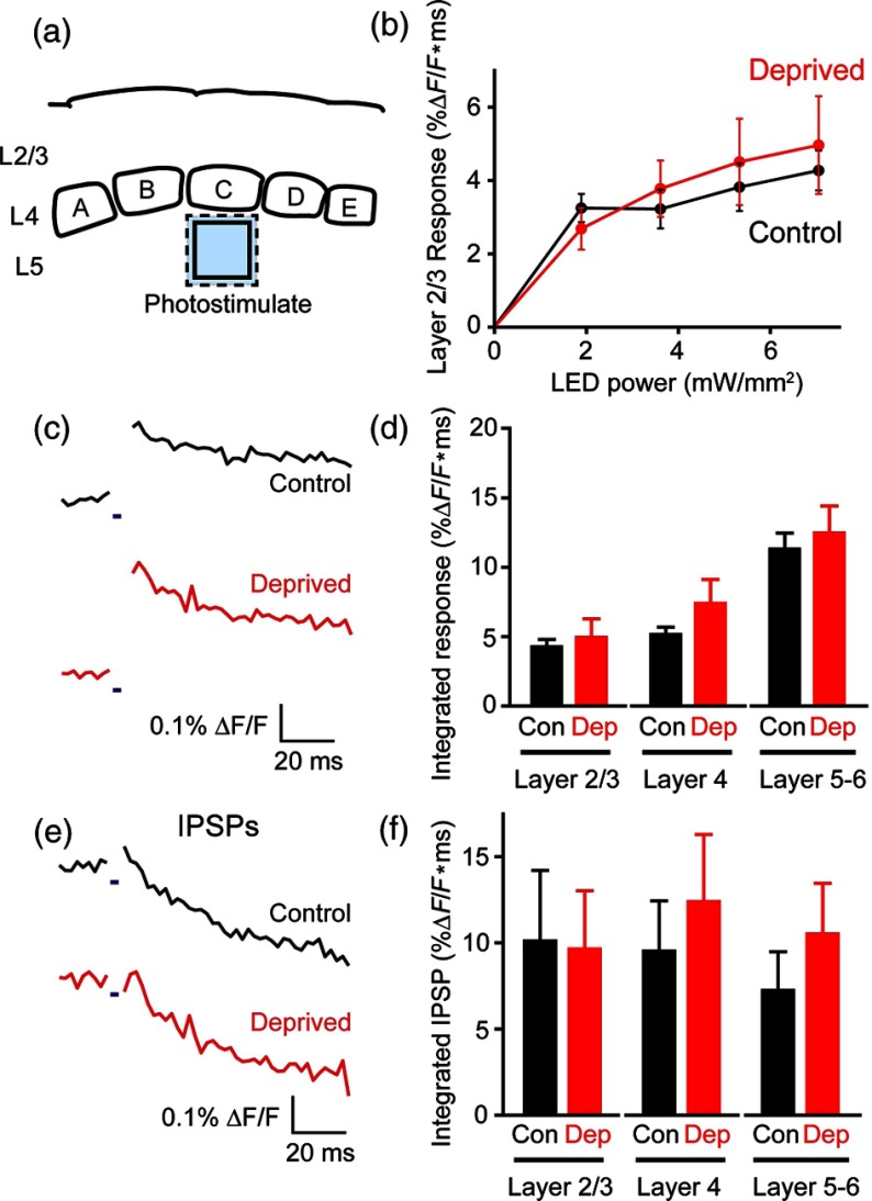
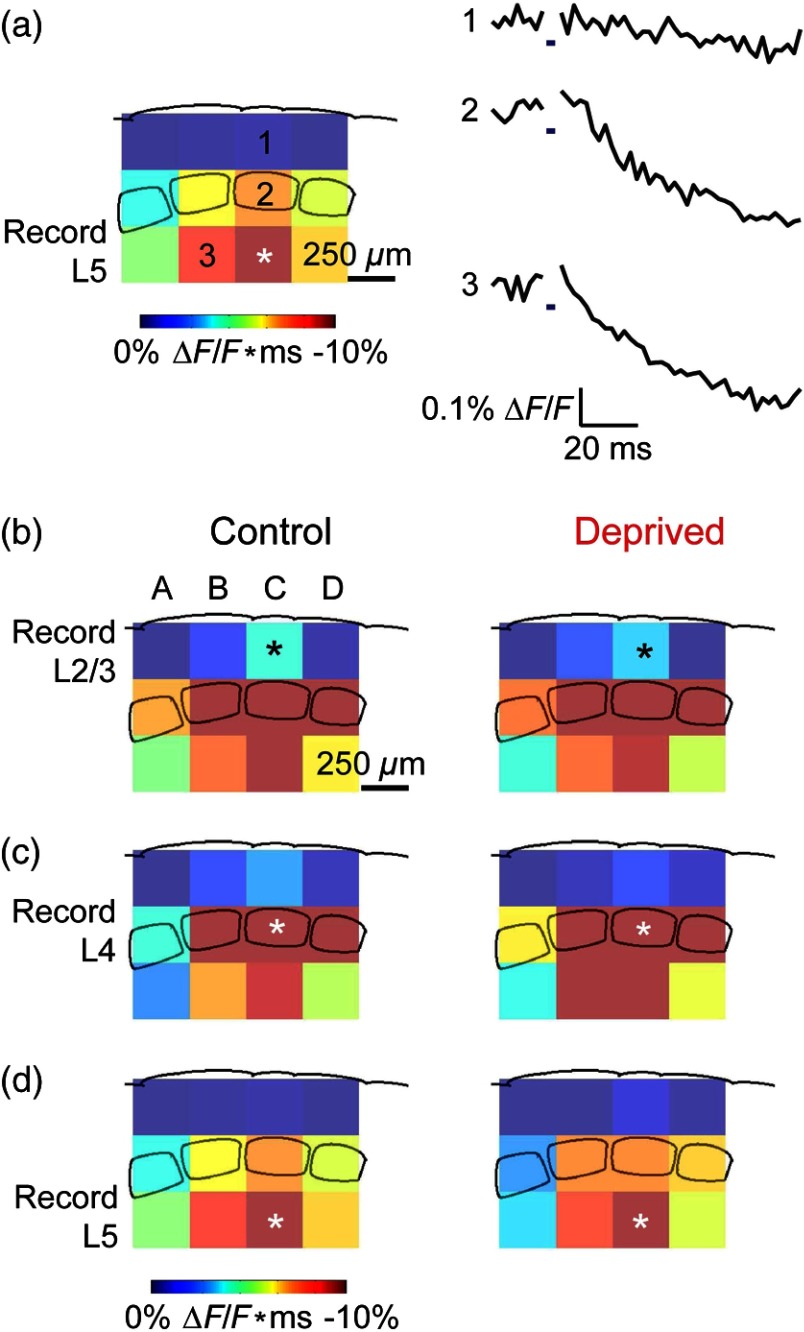
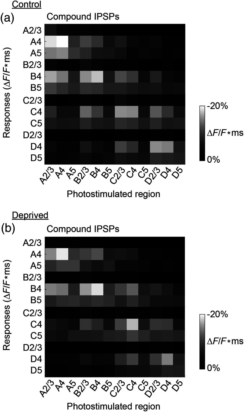
We describe an experimental approach that uses light to both control and detect neuronal activity in mouse barrel cortex slices: blue light patterned by a digital micromirror array system allowed us to photostimulate specific layers and columns, while a red-shifted voltage-sensitive dye was used to map out large-scale circuit activity. We demonstrate that such all-optical mapping can interrogate various circuits in somatosensory cortex by sequentially activating different layers and columns. Further, mapping in slices from whisker-deprived mice demonstrated that chronic sensory deprivation did not significantly alter feedforward inhibition driven by layer 5 pyramidal neurons. Further development of voltage-sensitive optical probes should allow this all-optical mapping approach to become an important and high-throughput tool for mapping circuit interactions in the brain.

Keywords: barrel cortex; channelrhodopsin; optogenetics; voltage-sensitive dye.


Figures

Fig. 1
Fig. 1
Expression of channelrhodopsin-2 (ChR2) in layer 5 pyramidal neurons in the barrel cortex of Thy1-ChR2 mice. (a) Expression of eYFP-tagged ChR2 (in green) in the cortex of homozygous Thy1-ChR2 line 18 mice. (b) Preferential expression of eYFP-tagged ChR2 in layer 5 pyramidal neurons in the cortex.
Fig. 2
Fig. 2
Light-evoked responses of multiple neuron types in barrel cortex slices from Thy1-ChR2 mice. (a) Action potential firing pattern (top trace) elicited in a layer 5 pyramidal cell in response to a 1 s duration depolarizing current pulse (lower trace). (b) Light-evoked responses produced by photostimulation (3 ms duration, at arrow). (c) Action potential firing pattern (top trace) elicited in a fast-spiking basket cell in response to a 1 s duration depolarizing current pulse (lower trace). (d) Light-evoked response produced by photostimulation (3 ms duration, at arrow). (e) Action potential firing pattern elicited in a nonfast-spiking interneuron in response to a 1 s duration depolarizing current pulse (lower trace). (f) Light-evoked depolarizing inhibitory postsynaptic potential (IPSP) produced by photostimulation (3 ms duration, at arrow).
Fig. 3
Fig. 3
Photostimulation of layer 5 neurons elicits postsynaptic responses in layer 2/3. (a) All-optical stimulation of neurons with 460 nm light-emitting diode (LED) light and recording of excitation by voltage-sensitive dye (VSD) fluorescence. The mosaic micromirror array was used to photostimulate specific regions of the slice. (b) Diagram depicting photostimulation of layer 5 pyramidal neurons in columns A to E is shown in the top-left panel. Other panels are VSD images showing the time-dependent spread of circuit activity from layers 5 to 2/3 in the somatosensory cortex. A 460 nm light flash (5 ms) was used to photostimulate layer 5 pyramidal neurons and images taken at respective durations after starting photostimulation are shown. Barrels are indicated in white to indicate the location of columns A to E. (c) VSD image taken at 17.6 ms after the onset of a 5 ms 460 nm light flash to stimulate layer 5 pyramidal neurons (indicated by the dotted area at location 2). (d) Postsynaptic responses measured in layer 2/3 were blocked with bath application of 6-cyano-7-nitroquinoxaline-2,3-dione (CNQX) (10μM). In these and all other traces, horizontal bars indicate the timing of photostimulation. Transient fluorescence increases caused by excitation of the VSD during photostimulation have been blanked out.
Fig. 4
Fig. 4
Photostimulation of layer 5 pyramidal neurons lead to intercolumnal spread of postsynaptic activity. (a) Diagram depicting arrangement for photostimulation of layer 5 pyramidal neurons in column C. (b) VSD image taken at 13.2 ms after the onset of a 3 ms 460 nm light flash to stimulate layer 5 pyramidal neurons (indicated by the dotted box at location 2), showing the spread of excitatory responses up to layer 2/3 and along layer 5. Barrels are indicated in white and lettered accordingly, showing the location of A to E columns. (c) Different peak latencies in layers 2/3 and 5 responses show that responses initiated in layer 5 spread toward layer 2/3. (d) VSD image taken at 13.2 ms after a 50μA electrical pulse was delivered to layer 5. Strong responses tended to spread upwards along the column. (e) Electrically stimulated slices show a similar pattern of peak latencies in layers 2/3 and 5, indicating that responses initiated in layer 5 spread toward layer 2/3.
Fig. 5
Fig. 5
All-optical mapping can be done at a higher resolution. (a) Input response map for layer 5 of column C of slices from a P113 mouse done at lower (12 photoactivation areas, top) and higher (54 photoactivation areas, bottom) resolution. Integrated responses elicited at the region denoted by an asterisk following photostimulation of various areas on the slice were color-coded accordingly. Input map responses were integrated from 7.4 to 25 ms after the onset of the light flash. (b) and (c) Averaged optical traces show responses measured in layer 5 (indicated by asterisk) when photostimulating in layer 5. A more refined response map can be obtained at higher resolution of photostimulation, showing differential amounts of light-evoked responses within a region that would otherwise be masked at a lower resolution. Traces are numbered as indicated on the map.
Fig. 6
Fig. 6
Chronic whisker deprivation did not significantly affect layer 5 pyramidal neuron-driven excitatory responses and feedback inhibition. (a) Diagram showing the photostimulation of layer 5 neurons in column C. The blue dotted box indicates the photostimulation area while the solid box indicates the area of interest where responses from (c) and (e) are taken from. (b) Similar input–output curves of layer 2/3 responses following photostimulation of layer 5 pyramidal neurons for control (black) and deprived (red) slices. Maximum LED power was used for photostimulation experiments in the rest of the study. Integrated layer 2/3 responses were integrated from 7.4 to 25 ms after the onset of the light flash. (c) Averaged optical traces of layer 5 responses following photostimulation of layer 5 pyramidal neurons for control (black) and deprived (red) slices. 3 ms photostimulation indicated by the black bar below the trace. Deprived responses were slightly larger than controls but not significantly so. (d) Histogram of excitatory responses at layers 2/3, 4, and 5 of column C following photostimulation of layer 5 in (a). Excitatory responses were not significantly different across all layers. Integrated responses at various layers were integrated from 7.4 to 25 ms after the onset of the light flash. (e) Averaged optical responses elicited at layer 5 following photostimulation of L5 pyramidal neurons in column C for control (black) and deprived (red) slices. 3 ms photostimulation indicated by the black bar below the trace. Subtracted compound IPSPs were similar between control and deprived slices. (f) Histogram of compound IPSP responses at layers 2/3, 4, and 5 following photostimulation of L5 pyramidal neurons in column C as in (a). Compound IPSP responses were not significantly different across all layers, suggesting that experience-dependent plasticity involves interneurons not driven by layer 5 pyramidal cells. Integrated compound IPSP responses at various layers were integrated from 11.8 to 51.4 ms after the onset of the light flash.
Fig. 7
Fig. 7
All-optical mapping of column C responses with ChR2 photostimulation and VSD imaging. (a) Averaged control input response map (left; n=6) for layer 5 of column C. Input map responses were integrated from 7.4 to 25 ms after the onset of the light flash. Averaged optical traces (right) showing responses measured in layer 5 (indicated by asterisk) when photostimulating layers 2/3 and 4 of column C and layer 5 of column B. Traces are numbered as indicated on the map. (b) to (d) Averaged input response maps (n=6) for column C, showing the strength of compound IPSP input from surrounding layers and columns of the slice. Position of asterisk indicates layer and column of area from which responses were calculated. Response maps show similar input patterns between controls and deprived slices.
Fig. 8
Fig. 8
Response matrices from high-throughput all-optical circuit mapping of the barrel cortex. (a) Averaged compound IPSP input responses of control slices (n=6), showing the strength of compound IPSP inputs from all columns and layers for all columns and layers. Responses elicited in various layers (2/3, 4, or 5) of different columns (A to D) were arranged along the y-axis and the region of photostimulation arranged along the x-axis as a response matrix. The strength of compound IPSP responses are color-coded in grayscale as indicated in the scale bar. (b) Averaged compound IPSP input responses of deprived slices (n=6), showing the strength of compound IPSP inputs from all columns and layers to all columns and layers. The pattern of responses is similar to the controls and responses were not significantly different in each region (two sample t tests, n=6).

Similar articles

Cited by

References

    1. Lustig B. R., et al. , “Voltage-sensitive dye imaging reveals shifting spatiotemporal spread of whisker-induced activity in rat barrel cortex,” J. Neurophysiol. 109, 2382–2392 (2013).JONEA410.1152/jn.00430.2012 - DOI - PMC - PubMed
    1. Petersen C. C., Sakmann B., “Functionally independent columns of rat somatosensory barrel cortex revealed with voltage-sensitive dye imaging,” J. Neurosci. 21, 8435–8446 (2001).JNRSDS - PMC - PubMed
    1. Laaris N., Keller A., “Functional independence of layer IV barrels,” J. Neurophysiol. 87, 1028–1034 (2002).JONEA4 - PMC - PubMed
    1. Laaris N., Carlson G. C., Keller A., “Thalamic-evoked synaptic interactions in barrel cortex revealed by optical imaging,” J. Neurosci. 20, 1529–1537 (2000).JNRSDS - PMC - PubMed
    1. Petersen C. C., Grinvald A., Sakmann B., “Spatiotemporal dynamics of sensory responses in layer 2/3 of rat barrel cortex measured in vivo by voltage-sensitive dye imaging combined with whole-cell voltage recordings and neuron reconstructions,” J. Neurosci. 23, 1298–1309 (2003).JNRSDS - PMC - PubMed