. 2014 Apr;1(1):014004.
doi: 10.1117/1.JMI.1.1.014004. Epub 2014 Apr 23.

Multiframe registration of real-time three-dimensional echocardiography time series

Affiliations

Multiframe registration of real-time three-dimensional echocardiography time series

Harriët W Mulder et al. J Med Imaging (Bellingham). 2014 Apr.

Abstract

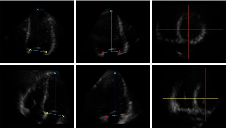
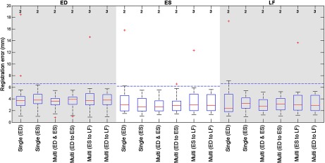
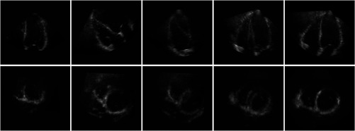
Mosaicing of real-time three-dimensional echocardiography (RT3-DE) images aims at extending the field-of-view of overlapping images. Currently available methods discard most of the temporal information available in the time series. We investigate the added value of simultaneous registration of multiple temporal frames using common similarity metrics. We combine RT3-DE images of the left and right ventricles by registration and fusion. The standard approach of registering single frames, either end-diastolic (ED) or end-systolic (ES), is compared with simultaneous registration of multiple time frames, to evaluate the effect of using the information from all images in the metric. A transformation estimating the protocol-specific misalignment is used to initialize the registration. It is shown that multiframe registration can be as accurate as alignment of the images based on manual annotations. Multiframe registration using normalized cross-correlation outperforms any of the single-frame methods. As opposed to expectations, extending the multiframe registration beyond simultaneous use of ED and ES frames does not further improve registration results.

Keywords: image mosaicing; intensity-based registration; multiframe registration; real-time three-dimensional echocardiography.


Figures

Fig. 1
Fig. 1
The annotated left ventricle (LV) data (upper row) and right ventricle (RV) data (lower row) at end diastole (ED); two-dimensional (2-D) slices from a RT3-DE data set. From left to right: Four-chamber view. Two-chamber view. Short-axis view, the directions of the four- and two-chamber views are depicted in yellow and red, respectively. The crosses indicate the position of the apex and the four points on the mitral valve annulus that were used for evaluation.
Fig. 2
Fig. 2
Boxplot showing the registration errors at end diastole (ED), end systole (ES) and the last time frame (LF) for the different registration strategies. NCC was used as metric. Multiframe registration with ED and ES frames results in the smallest deviation in the registration errors. At the top the number of data sets with a registration error over 20 mm is given. The horizontal (dashed) lines give the maximal interobserver transformation distance, or ITD, for the different temporal frames.
Fig. 3
Fig. 3
The four-chamber view (top row) and short-axis view (lower row) at ED; 2-D slices of a 3-D volume data set are shown. From left to right: LV image. RV image. Fused LV and RV images (alignment of image axes). Fused LV and RV images (LV data is transformed by the initial transformation). Fused LV and RV images after successful registration (NCC, multiframe registration including ED and ES frames). Mean-intensity fusion was used for image fusion.

References

    1. Burri M. V., et al. , “Review of novel clinical applications of advanced, real-time, 3-dimensional echocardiography,” Transl. Res. 159(3), 149–164 (2012).TRRECL10.1016/j.trsl.2011.12.008 - DOI - PubMed
    1. Wachinger C., Wein W., Navab N., “Registration strategies and similarity measures for three-dimensional ultrasound mosaicing,” Acad. Radiol. 15(11), 1404–1415 (2008).10.1016/j.acra.2008.07.004 - DOI - PubMed
    1. Szmigielski C., et al. , “Real-time 3D fusion echocardiography,” JACC Cardiovasc. Imaging 3(7), 682–690 (2010).10.1016/j.jcmg.2010.03.010 - DOI - PubMed
    1. Rajpoot K., et al. , “Multiview fusion 3-D echocardiography: improving the information and quality of real-time 3-D echocardiography,” Ultrasound Med. Biol. 37(7), 1056–1072 (2011).USMBA310.1016/j.ultrasmedbio.2011.04.018 - DOI - PubMed
    1. Rajpoot K., et al. , “The evaluation of single-view and multi-view fusion 3D echocardiography using image-driven segmentation and tracking,” Med. Image Anal. 15(4), 514–528 (2011).MIAECY10.1016/j.media.2011.02.007 - DOI - PubMed