. 2014 Oct;1(3):033502.
doi: 10.1117/1.JMI.1.3.033502. Epub 2014 Nov 3.

Quantitative simultaneous positron emission tomography and magnetic resonance imaging

Affiliations

Quantitative simultaneous positron emission tomography and magnetic resonance imaging

Jinsong Ouyang et al. J Med Imaging (Bellingham). 2014 Oct.

Abstract

Simultaneous positron emission tomography and magnetic resonance imaging (PET-MR) is an innovative and promising imaging modality that is generating substantial interest in the medical imaging community, while offering many challenges and opportunities. In this study, we investigated whether MR surface coils need to be accounted for in PET attenuation correction. Furthermore, we integrated motion correction, attenuation correction, and point spread function modeling into a single PET reconstruction framework. We applied our reconstruction framework to in vivo animal and patient PET-MR studies. We have demonstrated that our approach greatly improved PET image quality.

Keywords: motion correction; point spread function modeling; positron emission tomography and magnetic resonance imaging; positron emission tomography attenuation correction; surface coils.


Figures

Fig. 1
Fig. 1
Phantom study using Siemens mMR surface coils. (a) An mMR surface coil array was placed on the top of a data spectrum anthropomorphic torso phantom, (b) positron emission tomography (PET) bias image, (c) PET bias profiles.
Fig. 2
Fig. 2
A coronal PET FDG slice reconstructed using (a) OP-OSEM and (b) PSF-OP-OSEM. PSF modeling yielded higher radiotracer thyroid uptake to background ratios (TBR).
Fig. 3
Fig. 3
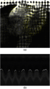
Magnetic resonance (MR) motion tracking in subject 2. (a) Tagged MR images with estimated motion fields (The displacement vector fields are depicted by the small yellow arrows). (b) Breathing navigator echoes (The bright dots indicate the navigator is in the reference respiratory phase).
Fig. 4
Fig. 4
Time-dependent PET attenuation map acquired on a subject. The reference attenuation map was directly obtained from the mMR scanner.
Fig. 5
Fig. 5
Sagittal MRI (a) and corresponding PET slices reconstructed using (b) respiratory gating (OP-OSEM “gated”) (c) no motion correction (OP-OSEM) (d) no motion correction with PSF modeling (PSF-OP-OSEM), (e) MR-based motion correction (MC-OP-OSEM) and (f) motion and PSF corrections (‘MC-PSF-OP-OSEM’). The gated volume was reconstructed using PET counts detected at end-exhalation (Approximately one-seventh of the total number of events). As can be seen, the proposed reconstruction methods (e and f) substantially improved hepatic lesion contrast and structure delineation as compared to the conventional uncorrected reconstruction method (c).
Fig. 6
Fig. 6
Heart short axis MRI (a) and corresponding PET slices reconstructed using (b) cardiac gating (OP-OSEM “gated”) (c) no motion correction (OP-OSEM) (d) no motion correction with PSF modeling (PSF-OP-OSEM), (e) MR-based motion correction (MC-OP-OSEM) and (f) motion and PSF corrections (“MC-PSF-OP-OSEM”). ROIs located on the myocardium for contrast calculations are shown in (a). The proposed reconstruction methods (e and f) substantially improved reconstructed myocardial contrast and wall delineation (see yellow arrows) as compared to the conventional uncorrected reconstruction method (c).

References

    1. Zaidi H., Montandon M. L., Slosman D. O., “Magnetic resonance imaging-guided attenuation and scatter corrections in three-dimensional brain positron emission tomography,” Med. Phys. 30(5), 937–948 (2003).MPHYA610.1118/1.1569270 - DOI - PubMed
    1. Zaidi H., Montandon M. L., Meikle S. R., “Strategies for attenuation compensation in neurological PET studies,” NeuroImage 34(2), 518–541 (2007).NEIMEF10.1016/j.neuroimage.2006.10.002 - DOI - PubMed
    1. Beyer T., et al. , “MR-based attenuation correction for torso-PET/MR imaging: pitfalls in mapping MR to CT data,” Eur. J. Nucl. Mol. Imaging 35(6), 1142–1146 (2008).EJNMA610.1007/s00259-008-0734-0 - DOI - PubMed
    1. Martinez-Moller A., et al. , “Tissue classification as a potential approach for attenuation correction in whole-body PET/MRI: evaluation with PET/CT data,” J. Nucl. Med. 50(4), 520–526 (2009).JNMEAQ10.2967/jnumed.108.054726 - DOI - PubMed
    1. Keereman V., et al. , “MRI-based attenuation correction for PET/MRI using ultrashort echo time sequences,” J. Nucl. Med. 51(5), 812–818 (2010).JNMEAQ10.2967/jnumed.109.065425 - DOI - PubMed