. 2014 Oct;1(3):035001.
doi: 10.1117/1.JMI.1.3.035001. Epub 2014 Oct 27.

Quantitative identification of magnetic resonance imaging features of prostate cancer response following laser ablation and radical prostatectomy

Affiliations

Quantitative identification of magnetic resonance imaging features of prostate cancer response following laser ablation and radical prostatectomy

Geert J S Litjens et al. J Med Imaging (Bellingham). 2014 Oct.

Abstract

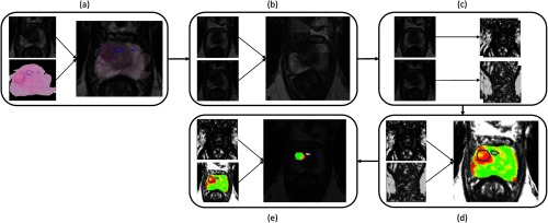
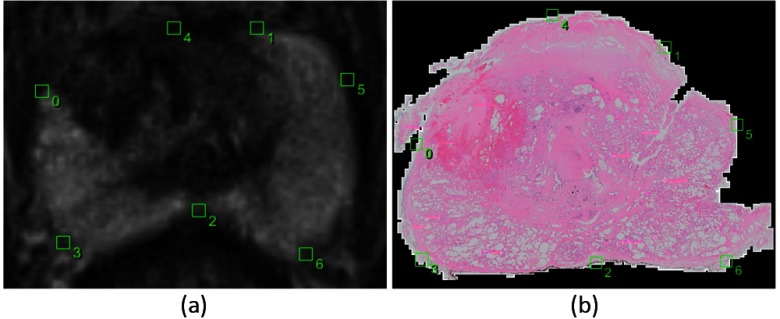
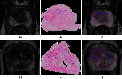
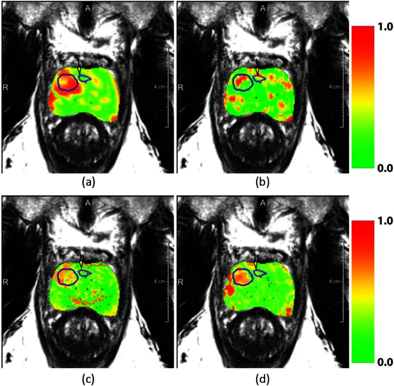
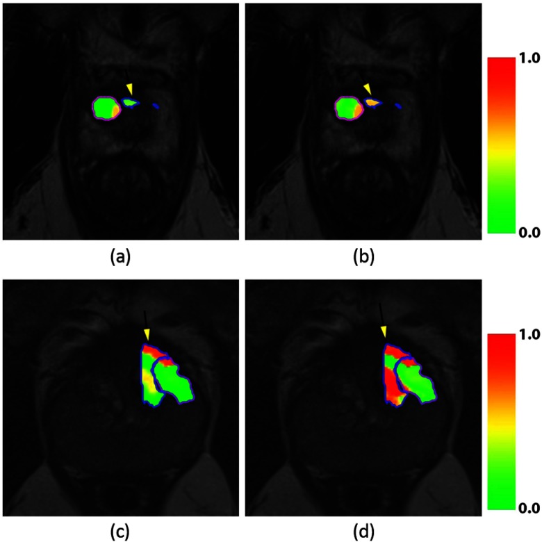
Laser interstitial thermotherapy (LITT) is a relatively new focal therapy technique for the ablation of localized prostate cancer. In this study, for the first time, we are integrating ex vivo pathology and magnetic resonance imaging (MRI) to assess the imaging characteristics of prostate cancer and treatment changes following LITT. Via a unique clinical trial, which gave us the availability of ex vivo histology and pre- and post-LITT MRIs, (1) we investigated the imaging characteristics of treatment effects and residual disease, and (2) evaluated treatment-induced feature changes in the ablated area relative to the residual disease. First, a pathologist annotated the ablated area and the residual disease on the ex vivo histology. Subsequently, we transferred the annotations to the post-LITT MRI using a semi-automatic elastic registration. The pre- and post-LITT MRIs were registered and features were extracted. A scoring metric based on the change in median pre- and post-LITT feature values was introduced, which allowed us to identify the most treatment responsive features. Our results show that (1) image characteristics for treatment effects and residual disease are different, and (2) the change of feature values between pre- and post-LITT MRIs can be a quantitative biomarker for treatment response. Finally, using feature change improved discrimination between the residual disease and treatment effects.

Keywords: laser ablation therapy; magnetic resonance imaging; prostate cancer; treatment response.


Figures

Fig. 1
Fig. 1
Flowchart detailing the process of (a) co-registration of histopathology and MRI using a semi-automated thin plate spline approach, (b) registration of pre- and post-LITT MRIs using affine registration with localized mutual information, (c) extraction of features (Table 1) and (d) calculation of relative feature change and finally (e) the clustering result to detect residual disease using fuzzy C-means clustering.
Fig. 2
Fig. 2
Example images of the corresponding points selected by the observer in the post-LITT axial T2-weighted MRI (a) and the corresponding histopathology slice (b). Points 0 and 1 indicate the ends of the peripheral zone, point 2 is the thinnest point of the peripheral zone, points 3 and 6 are the thickest parts and point 4 is the top part of the prostate when drawing a line from point 2 through the urethra to the top of the prostate. Point 5 was added due to the tear in the tissue.
Fig. 3
Fig. 3
Example images of the post-LITT MRI (a and d) and H and E stained prostatectomy slides (b and e) and the result of the subsequent MRI/histology registration (c and f). Ablated area in purple, residual disease in blue.
Fig. 4
Fig. 4
Example images of the pre-LITT axial T2-weighted MRI (a), post-LITT MRI (b), and the subsequent affine registration result in a checkerboard form (c). Registration was performed using localized mutual information as a metric.
Fig. 5
Fig. 5
Overlays of normalized feature change between pre- and post-treatment MRIs. Figures (a), (b), (c), and (d) represent the features Kep, Haralick correlation, (Pseudo)T2-Map, and Ve respectively. In the overlay, red indicates areas of high relative change and green of low relative change. The successfully ablated area is indicated in a blue contour and the purple contour indicates residual disease. The residual disease area is also indicated with a yellow arrow.
Fig. 6
Fig. 6
Relative change in feature value between residual disease (red, top bars) and the successfully ablated area (blue, bottom bars) for the top 10 scoring features (top to bottom). Almost all features are either pharmacokinetic (1, 5, and 10) or textural in nature (2, 3, 6, 7, 8, and 9). Furthermore, although texture features show more change in successfully ablated tissue relative to the pharmacokinetic features, they also show large change in the residual disease.
Fig. 7
Fig. 7
Likelihood heat maps for the fuzzy C-means clustering separating treatment effects and residual disease. Patient 1 (a and b) and patient 3 (c and d) are shown. Figures (a and c) show the results obtained when only including post-LITT MRI feature, whereas figures (b and d) show the improvement obtained by incorporating the 10 highest scoring features (Table 2). Residual disease in blue, ablated area in purple. The residual disease area is also indicated with a yellow arrow.

References

    1. Hegarty J., et al. , “Radical prostatectomy versus watchful waiting for prostate cancer,” Cochrane Database Syst. Rev. (11), CD006590 (2010). - PubMed
    1. Rosenkrantz A. B., et al. , “Role of MRI in minimally invasive focal ablative therapy for prostate cancer,” AJR Am. J. Roentgenol. 197(1), W90–W96 (2011).AJROAM10.2214/AJR.10.5946 - DOI - PubMed
    1. Oto A., et al. , “MR imaging-guided focal laser ablation for prostate cancer: phase I trial,” Radiology 267, 932–940 (2013).RADLAX10.1148/radiol.13121652 - DOI - PubMed
    1. Ahmed H. U., et al. , “Do low-grade and low-volume prostate cancers bear the hallmarks of malignancy?,” Lancet Oncol. 13, e509–e517 (2012).LOANBN10.1016/S1470-2045(12)70388-1 - DOI - PubMed
    1. Roberts H. R., et al. , “Dynamic magnetic resonance control of interstitial laser photocoagulation therapy of colorectal hepatic metastases,” Lancet 343, 1221 (1994).LANCAO10.1016/S0140-6736(94)92425-2 - DOI - PubMed