

Mammographic density measurements are not affected by mammography system
- PMID: 26158085
- PMCID: PMC4478994
- DOI: 10.1117/1.JMI.2.1.015501
Mammographic density measurements are not affected by mammography system
Abstract
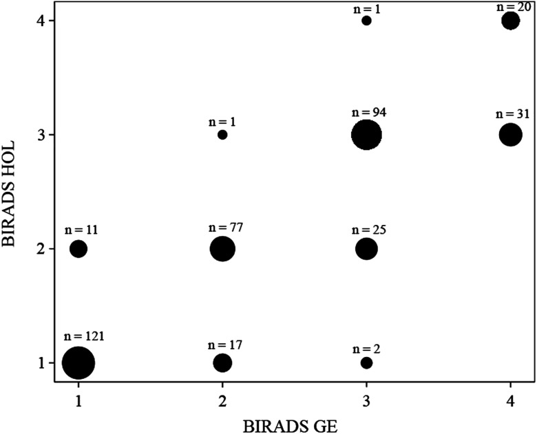
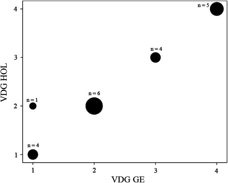
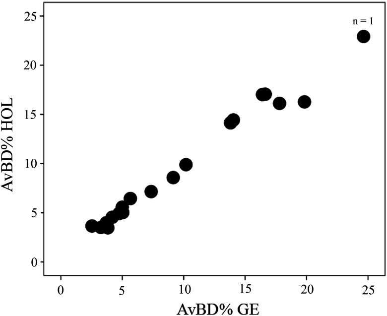
Mammographic density (MD) is a significant risk factor for breast cancer and has been shown to reduce the sensitivity of mammography screening. Knowledge of a woman's density can be used to predict her risk of developing breast cancer and personalize her imaging pathway. However, measurement of breast density has proven to be troublesome with wide variations in density recorded using radiologists' visual Breast Imaging Reporting and Data System (BIRADS). Several automated methods for assessing breast density have been proposed, each with their own source of measurement error. The use of differing mammographic imaging systems further complicates MD measurement, especially for the same women imaged over time. The purpose of this study was to investigate whether having a mammogram on differing manufacturer's equipment affects a woman's MD measurement. Raw mammographic images were acquired on two mammography imaging systems (General Electric and Hologic) one year apart and processed using VolparaDensity™ to obtain the Volpara Density Grade (VDG) and average volumetric breast density percentage (AvBD%). Visual BIRADS scores were also obtained from 20 expert readers. BIRADS scores for both systems showed strong positive correlation ([Formula: see text]; [Formula: see text]), while the VDG ([Formula: see text]; [Formula: see text]) and AvBD% ([Formula: see text]; [Formula: see text]) showed stronger positive correlations. Substantial agreement was shown between the systems for BIRADS ([Formula: see text]; [Formula: see text]), however, the systems demonstrated an almost perfect agreement for VDG ([Formula: see text]; [Formula: see text]).
Keywords: Breast Imaging Reporting and Data System; General Electric; Hologic; Volpara; mammographic density.
Figures
Similar articles
-
Mammographic Breast Density Assessment Using Automated Volumetric Software and Breast Imaging Reporting and Data System (BIRADS) Categorization by Expert Radiologists.Acad Radiol. 2016 Jan;23(1):70-7. doi: 10.1016/j.acra.2015.09.011. Epub 2015 Oct 26. Acad Radiol. 2016. PMID: 26514436
-
Inter-observer variability in mammographic density assessment using Royal Australian and New Zealand College of Radiologists (RANZCR) synoptic scales.J Med Imaging Radiat Oncol. 2016 Jun;60(3):329-36. doi: 10.1111/1754-9485.12451. Epub 2016 Apr 5. J Med Imaging Radiat Oncol. 2016. PMID: 27059785
-
Studying the association between longitudinal mammographic density measurements and breast cancer risk: a joint modelling approach.Breast Cancer Res. 2023 Jun 9;25(1):64. doi: 10.1186/s13058-023-01667-8. Breast Cancer Res. 2023. PMID: 37296473 Free PMC article.
-
Reporting breast density on chest computed tomography.Transl Breast Cancer Res. 2023 Jul 30;4:24. doi: 10.21037/tbcr-23-36. eCollection 2023. Transl Breast Cancer Res. 2023. PMID: 38751487 Free PMC article. Review.
-
Abnormal mammographic findings with short-interval follow-up recommendation.Clin Breast Cancer. 2005 Aug;6(3):235-9. doi: 10.3816/CBC.2005.n.025. Clin Breast Cancer. 2005. PMID: 16137434 Review.
Cited by
-
Age-related change in mammographic breast density of women without history of breast cancer over a 10-year retrospective study.PeerJ. 2023 Feb 14;11:e14836. doi: 10.7717/peerj.14836. eCollection 2023. PeerJ. 2023. PMID: 36815981 Free PMC article.
-
A practical work around for breast density distribution discrepancies between mammographic images from different vendors.Eur Radiol. 2025 Aug;35(8):4885-4892. doi: 10.1007/s00330-025-11383-w. Epub 2025 Jan 31. Eur Radiol. 2025. PMID: 39890617 Free PMC article.
-
Mammographic density changes during neoadjuvant breast cancer treatment: NeoDense, a prospective study in Sweden.Breast. 2020 Oct;53:33-41. doi: 10.1016/j.breast.2020.05.013. Epub 2020 Jun 4. Breast. 2020. PMID: 32563178 Free PMC article.
-
Mammographic density assessed on paired raw and processed digital images and on paired screen-film and digital images across three mammography systems.Breast Cancer Res. 2016 Dec 19;18(1):130. doi: 10.1186/s13058-016-0787-0. Breast Cancer Res. 2016. PMID: 27993168 Free PMC article.
-
Breast parenchymal patterns in processed versus raw digital mammograms: A large population study toward assessing differences in quantitative measures across image representations.Med Phys. 2016 Nov;43(11):5862. doi: 10.1118/1.4963810. Med Phys. 2016. PMID: 27806604 Free PMC article.
