. 2015 Jul 9:6:7670.
doi: 10.1038/ncomms8670.

A palette of fluorescent proteins optimized for diverse cellular environments

Affiliations

A palette of fluorescent proteins optimized for diverse cellular environments

Lindsey M Costantini et al. Nat Commun. .

Abstract

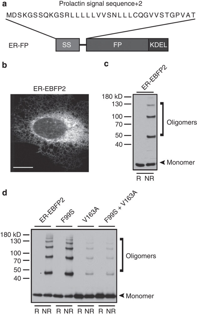
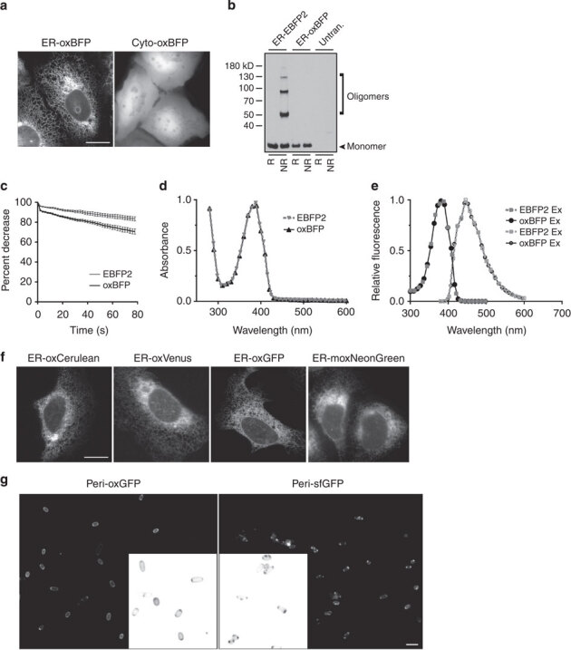
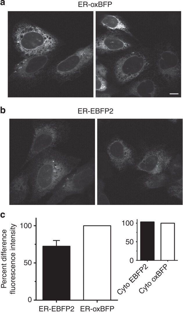
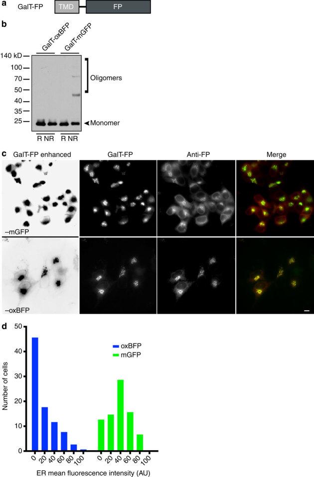
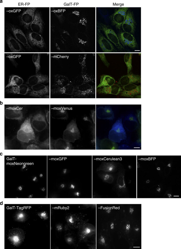
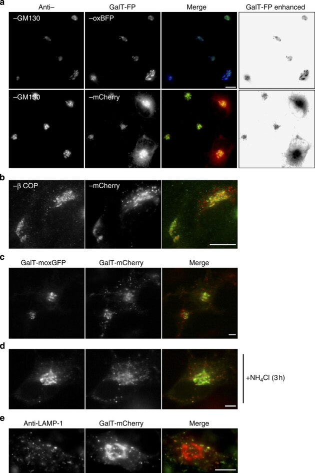
To perform quantitative live cell imaging, investigators require fluorescent reporters that accurately report protein localization and levels, while minimally perturbing the cell. Yet, within the biochemically distinct environments of cellular organelles, popular fluorescent proteins (FPs), including EGFP, can be unreliable for quantitative imaging, resulting in the underestimation of protein levels and incorrect localization. Specifically, within the secretory pathway, significant populations of FPs misfold and fail to fluoresce due to non-native disulphide bond formation. Furthermore, transmembrane FP-fusion constructs can disrupt organelle architecture due to oligomerizing tendencies of numerous common FPs. Here, we describe a powerful set of bright and inert FPs optimized for use in multiple cellular compartments, especially oxidizing environments and biological membranes. Also, we provide new insights into the use of red FPs in the secretory pathway. Our monomeric 'oxFPs' finally resolve long-standing, underappreciated and important problems of cell biology and should be useful for a number of applications.


Conflict of interest statement

A provisional patent application has been filed covering part of the results described in this manuscript (L.M.C. and E.L.S). Albert Einstein College Medicine and E.L.S. have licensed technology described in this manuscript to Lucigen Corp. The remaining authors declare no competing financial interests.

Figures

Figure 1
Figure 1. ER-localized EBFP2 forms non-native disulphide bonds.
(a) Schematic of ER-localization FP (ER–FP), containing prolactin signal sequence (SS) and KDEL retrieval motif. (b) Representative image of transiently transfected U-2 OS cells expressing ER–EBFP2; scale bar, 10 μm. (c) Immunoblot with reducing (R, +DTT) and nonreducing (NR, −DTT) conditions illustrate the tendency of ER–EBFP2 to oligomerize under NR conditions. The lower molecular weight band denotes expected molecular weight of monomeric EBFP2 (∼25 kD). (d) Superfolder and cycle-3 mutations do not protect EBFP2 cysteine residues from inappropriate disulphide bond formation. Immunoblot of ER–EBFP2, with superfolder mutations (S30R, Y39N, N105T, Y145F, I171V) and FP versions including cycle-3 GFP mutations (F99S, V163A) forms higher molecular weight oligomers under NR conditions.
Figure 2
Figure 2. OxFPs are fluorescent.
(a) Representative image of ER–oxBFP and Cyto-oxBFP expressing U-2 OS cell; scale bar, 10 μm. (b) Immunoblot of cells transfected with ER–EBFP2 or –oxBFP. Under nonreducing conditions, the optimized, cysteine-less oxBPF does not form inappropriate disulphide bonds; untran., untransfected. (c) oxBFP (black line) maintains moderately comparable photostability properties under standard imaging conditions compared with EBFP2 (grey line). Optimized oxBFP variant (black data points) has comparable spectral characteristics to parental EBFP2 variant (grey data points). (d) Absorbance measurements and (e) fluorescence excitation (closed data points) and emission (open data points). (f) Representative images of ER–oxCerulean, –oxVenus, –oxGFP or moxNeonGreen expressing U-2 OS cells. Scale bar, 10 μm. (g) oxGFP fluoresces homogenously throughout the periplasm of gram-negative bacteria (CodonPlus competent, BL21 RP). Cells were induced for 1 h with IPTG and then imaged. Inset thumbnails are enlarged and inverted for detail. Scale bar, 1 μm.
Figure 3
Figure 3. ER-localized oxBFP has greater fluorescence intensity.
Representative images taken with identical imaging conditions of live U-2 OS cells transiently transfected (16 h post transfection) with (a) ER–oxBFP or (b) ER–EBFP2 illustrate the higher fluorescence level of the cysteine-less variant; scale bar, 10 μm. (c) Quantification of the percent difference of relative mean fluorescent intensity of the ER fluorescence signal ER–EBFP2 has an ∼27% decrease in brightness, total n=250 cells (ER–EBFP2 73 cells, ER–oxBFP 72 cells, Cyto-EBFP2 43 cells and Cyto-oxBFP 62 cells). Error bars signify s.e.m. for data collected from three experimental replicates.
Figure 4
Figure 4. Golgi complex membrane-localized mGFP forms inappropriate disulphide bonds and is inefficiently trafficked to the GC.
(a) Schematic of GC-localized FP (GalT–FP) containing GalT transmembrane domain upstream of FP. (b) Immunoblot revealed the tendency of GalT–mGFP to form oligomers under NR conditions. Optimized, cysteine-less GalT–oxBFP does not form inappropriate disulphide bonds. (c) Representative images of HeLa cells transiently transfected with GalT–mGFP or –oxBFP. Immunofluorescence with anti-GFP revealed a significant fluorescently undetectable pool of GalT–mGFP in the ER. ER labelling by the FP is digitally enhanced with Levels tool in Photoshop in far left panels. Note that weak ER is apparent in all of the oxBFP expressing cells, but rarely observed in mGFP expressing cells. Scale bar, 10 μm. (d) Distribution of the ER fluorescence intensity values (mean fluorescence intensities of regions of anti-GFP staining proximal to the GC). n≥80 cells collected from 11 to 13 fields per construct.
Figure 5
Figure 5. Golgi complex-localized membrane fusions with oxBFP exhibit significant differences relative to red FP fusions.
Representative image of (a) HeLa cells transiently co-expressing ER–oxGFP and GalT–oxBFP or –mCherry. (b) Live cells expressing GalT–moxVenus and ER–moxCerulean3 are readily distinguishable and thus represent a useful pair of FPs for two-colour imaging in the secretory pathway of live cells. (c) Transiently transfected HeLa cells expressing GalT–moxNeonGreen, –oxGFP, –moxCerulean3, –moxBFP. (d) Live cells expressing GalT–TagRFP or GalT–mRuby2 exhibit both the GC and bright puncta throughout the cell, while GalT–FusionRed appears to primarily localize to GC structures. Scale bar, 10 μm.
Figure 6
Figure 6. GalT–mCherry puncta localize to lysosomes.
(a) Hela cells expressing GalT–oxBFP (upper panels) or GalT–mCherry (lower panels) were fixed and stained with the GC marker anti-GM130. Co-localization was observed for GalT–oxBFP, but cells expressing moderate levels of GalT–mCherry exhibit both GC co-localization and bright puncta throughout the cell. (b) GalT–mCherry expressing HeLa cells fixed and stained for the GC and intermediate compartment marker β COP. The red puncta do not co-localize with the green β COP puncta. (c,d) HeLa cells co-expressing GalT–moxGFP and GalT–mCherry show co-localization in the GC structure, but GalT–moxGFP does not localize to red puncta. (d) Pretreatment with NH4Cl for 3 h leads to substantial co-localization of GalT–moxGFP with red puncta. (e) GalT–mcherry expressing HeLa cells were fixed and stained for the lysosomal marker anti-LAMP1. Several red puncta co-localize with the LAMP1 positive structures. Scale bar, 10 μm.

References

    1. Prasher DC, Eckenrode VK, Ward WW, Prendergast FG, Cormier MJ. Primary structure of the Aequorea victoria green-fluorescent protein. Gene. 1992;111:229–233. - PubMed
    1. Rizzo MA, Davidson MW, Piston DW. Fluorescent protein tracking and detection: fluorescent protein structure and color variants. Cold Spring Harb. Protoc. 2009;2009:pdb.top63. - PubMed
    1. Shaner NC, Patterson GH, Davidson MW. Advances in fluorescent protein technology. J. Cell Sci. 2007;120:4247–4260. - PubMed
    1. Miyawaki A, Shcherbakova DM, Verkhusha VV. Red fluorescent proteins: chromophore formation and cellular applications. Curr. Opin. Struct. Biol. 2012;22:679–688. - PMC - PubMed
    1. Shcherbakova DM, Subach OM, Verkhusha VV. Red fluorescent proteins: advanced imaging applications and future design. Angew. Chem. Int. Ed. 2012;51:10724–10738. - PMC - PubMed

Publication types