. 2015 Jul 9:6:7620.
doi: 10.1038/ncomms8620.

PTEN regulates DNA replication progression and stalled fork recovery

Affiliations

PTEN regulates DNA replication progression and stalled fork recovery

Jinxue He et al. Nat Commun. .

Abstract

Faithful DNA replication is a cornerstone of genomic integrity. PTEN plays multiple roles in genome protection and tumour suppression. Here we report on the importance of PTEN in DNA replication. PTEN depletion leads to impairment of replication progression and stalled fork recovery, indicating an elevation of endogenous replication stress. Exogenous replication inhibition aggravates replication-originated DNA lesions without inducing S phase arrest in cells lacking PTEN, representing replication stress tolerance. iPOND analysis reveals the physical association of PTEN with DNA replication forks and PTEN-dependent recruitment of Rad51. PTEN deletion results in Rad51 dissociation from replication forks. Stalled replication forks in Pten-null cells can be reactivated by ectopic Rad51 or PTEN, the latter facilitating chromatin loading of Rad51. These data highlight the interplay of PTEN with Rad51 in promoting stalled fork restart. We propose that loss of PTEN may initiate a replication stress cascade that progressively deteriorates through the cell cycle.


Conflict of interest statement

Competing financial interests

The authors declare no competing financial interests.

Figures
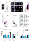
Figure 1
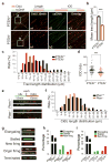
Figure 1. PTEN depletion induces FANCD2-associated anaphase bridges and premature S-phase exit
(a) Anaphase errors increase in PTEN-depleted cells. HeLa cells with or without PTEN shRNA (n=128 and 103 respectively) were examined by immunofluorescence for mitotic defects. Percentages of mitotic cells with anaphase bridges and micronuclei (arrowheads) are summarized in the histogram with represent DAPI images shown on the left. Data are presented as means±SEM and analyzed by unpaired t test. *, p<0.05. (b) Specification of pre-mitotic errors originating from DNA replication barrier. Immunofluorescence of FANCD2 was employed for labeling damaged DNA in mitosis reflective of replication defects. CENPA and DAPI were used to indicate centromeres and chromosomes. Top panel, anaphase and telophase cells without prominent FANCD2 foci. Mid and bottom panels, cells in anaphase or telophase with pronounced FANCD2 foci. Brackets, paired foci; Arrowheads, single or accumulated foci; Scale bar, 5 μm. (c) Frequencies of FANCD2-associated ana-telophase bridges summarized from FANCD2 immunofluorescence in cells with or without shPTEN in the presence and absence of aphidicolin (APH) treatment (0.1μM, 24 h). Data are presented as means±SEM (n>50 for each column) and analyzed by ANOVA with Turkey’s multiple comparisons test. *, p<0.05; **, p<0.01. (d) APH-treated HeLa cells with or without shPTEN were immunostained as in (b). Mitotic cells were analyzed for FANCD2 foci numbers and shown in a scatter plot. Data were processed by ANOVA and statistical significance was determined by Turkey’s multiple comparisons test. *, p<0.05; ***, p<0.001. (e) The distribution frequency of FANCD2 foci number in HeLa cells with or without shPTEN in the absence of APH treatment. (f) PTEN+/+ and PTEN−/− DLD-1 cells were treated with 1μM APH for 0, 2 or 6 h, prior to BrdU labeling and propidium iodide (PI) staining for flow cytometry analysis of cell cycle distribution. S phase was divided into three sub-S phases (early-S1, mid-S2 and late-S3) as indicated. (g and h) Summaries of cell cycle distribution in different phases (G0/G1, S and G2/M, h) or sub-S phases (S1+S2 and S3, h) respectively.
Figure 2
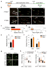
Figure 2. Replication fork progression is impaired in the absence of PTEN
(a) PTEN+/+ and PTEN−/− DLD-1 cells were pulse-labeled with CldU and subjected to single DNA fiber analysis. CldU tracts were visualized in red and ssDNA in green. Above the images is a schematic of the assay and data shown are from a single representative experiment out of three repeats. Scale bar, 25 μm. (b) Summary of mean CldU tract length in PTEN-proficient (n=302) and -deficient (n=301) cells. Data are presented as means±SEM and analyzed by unpaired t test. ***, p<0.001. (c) CldU fiber tract length distributions in PTEN+/+ and PTEN−/− cells. 1μm = 2 kb. (d) Distributions of the inter-origin distances (IOD) in PTEN+/+ and PTEN−/− cells. IOD was determined by measuring the distance between two identified replication initiation origins, as indicated by the sketch in (a). Bars and error bars represent means±SEM (n>75 in each cell group); ***, p<0.001. (e and f) Pten+/+ and Pten−/− MEFs were sequentially pulse-labeled with 50μM IdU and 50μM CldU, each for 15min. Green tracts, IdU; red tracts, CldU. A sketch delineating experimental design and representative images of dual-labeled fibers are shown in (e). Second-labeled CldU tract length distributions in Pten+/+ (n=302) and Pten−/− (n=300) cells are shown in (f). (g) Examples of fiber tract types representing different classes of replication structures. (h and i) Replication profiles of PTEN-proficient and -deficient MEFs (h) and DLD-1 cells (i) are presented by scoring relative frequencies of elongating fibers versus stalled or new-fired fibers. Minimums of 300 fibers were scored in each cell line.
Figure 3
Figure 3. PTEN is required for restart of DNA replication at stalled forks
(a) A schematic protocol of dual labeling DNA fiber assay for evaluation of replication restart following HU or APH-induced fork stalling. (b) PTEN+/+ and PTEN−/− DLD-1 cells were pre-labeled with CldU and then treated with 2mM HU for 2 h, prior to post-labeling with IdU for monitoring replication recovery. Selected areas are magnified for visual enhancement of stalled CldU tracts and gaps between CldU and IdU tracts in PTEN-null cells. Scale bar, 10 μm. (c and d) Summary of stalled fork frequencies (n>300) and gap lengths (n>200) between stalled sites and their corresponding re-initiation sites. Data are presented as means±SEM and analyzed by unpaired t test. **, p<0.01; ***, p<0.001. (e) Summary of DNA fiber restart assay in PTEN+/+ (n=284) and PTEN−/− (n=336) cells following APH (3μM) treatment. Data were scored and analyzed as in (d). **, p<0.01. (f) Pten+/+ and Pten−/− MEFs were pulse-labeled with CldU and subsequently treated with HU (2mM) or APH (3μM) for 2 h, followed by IdU pulse labeling to monitor replication recovery. A minimum of 200 fiber units was scored for each criterion. Data are presented as means±SEM and analyzed by unpaired t test. *, p<0.05. (g) BrdU incorporation analysis in response to APH treatment. HeLa cells with and without shRNA-mediated PTEN knockdown were subjected to 3μM APH treatment for different time periods as indicated, prior to BrdU pulse labeling for 15 min. Cells were then fixed for immunofluorescence of BrdU. Scale bar, 25 μm. (h) Immunofluorescence images from (g) were analyzed for BrdU intensity and summarized as mean intensity per cell (n>150). Data are presented as means±SEM in relative arbitrary unit and analyzed by unpaired t test. n.s., no significance; ***, p<0.001. (i) Pten+/+ and Pten−/− MEFs were treated with 3μM APH for 30min followed by BrdU pulse labeling. Immunofluorescence intensities of BrdU were scored before and after APH treatment. Data were analyzed by ANOVA and statistical significance was determined by Turkey’s multiple comparison tests. n.s., no significance; **, p<0.01.
Figure 4
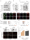
Figure 4. PTEN maintains replication proteins on chromatin for stalled fork recovery
(a) Cellular fractionation was performed in PTEN-proficient or -deficient DLD-1 cells to separate cytoplasm soluble fraction, nuclear soluble fraction and chromatin fraction, as described in Materials and Methods. Protein lysates from different fractions were immunoblotted with Rad51, PCNA and Chk1 antibodies. Tubulin was used as a loading control. (b) PTEN+/+ DLD-1 cells were subjected to TSA treatment (10uM, 12 h) followed by chromatin isolation for Western analysis of Rad51, PCNA and Chk1 respectively. (c) Levels of Rad51, PCNA and Chk1 were evaluated with immunoblotting using chromatin fractions from PTEN+/+ and PTEN−/− DLD-1 cells in the presence and absence of HU treatment. (d and e) APH-treated PTEN knockdown and control cells were pulse labeled with BrdU followed by Rad51 and PCNA immunofluorescence. Scale bar, 25 μm. (f and g) Immunofluorescent intensity of Rad51 (in d) and PCNA (in e) was quantified and summarized in BrdU positive and negative categories. One-way ANOVA was used for statistical analysis and Bonferroni’s multiple comparison tests were used for evaluation of statistical significance. **, p<0.01; ***, p<0.001. n.s. not significant. (h) Western blots to show overexpression of Rad51 or FLAG-tagged Chk1 in Pten−/− MEFs. (i) Pten+/+ and Pten−/− MEFs were pre-labeled with CldU and then treated with 0.5mM HU for 2 h, prior to post-labeling with IdU for monitoring replication recovery. Shown is representative DNA fiber images of indicated samples. Scale bar, 25 μm. (j) Summary of DNA stalled fork restart in Pten+/+ and Pten−/− cells with and without ectopic Rad51 or Chk1 following HU treatment (n>1000 in each sample). Data were analyzed with one-way ANOVA followed by Turkey’s multiple comparison tests. *, p<0.05; **, p<0.01, n.s., not significant.
Figure 5
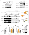
Figure 5. PTEN is physically associated with DNA replication forks and PTEN recruits Rad51 on chromatin for stalled fork recovery
(a) Chromatin was isolated from PTEN+/+ and PTEN−/− DLD-1 cells in the presence and absence of 2mM HU for indicated time periods, prior to PTEN immunoblotting. (b) Cell lysates were prepared from PTEN+/+ DLD-1 cells following sonication-induced chromatin lysis. Cells with and without HU treatment (2mM, 2 h) were subjected to PTEN immunoprecipitation followed by immunoblotting of Rad51 and Chk1. (c) PTEN+/+ and PTEN−/− HCT116 cells were labeled with EdU and treated with 2mM HU. Nascent DNA was conjugated with biotin for to iPOND analysis as described in Materials and Methods. DNA replication fork-associated PTEN, Rad51 and Chk1 were detected by Western blotting. Histone H3 was used a loading control. (d) A schematic model to show formation of the PTEN-Rad51-Chk1 complex on DNA replication forks in normal conditions and Rad51 dissociation (Chk1 may retain) following PTEN depletion. (e) PTEN overexpression in Pten−/− MEFs. (f and g) Pten−/− MEFs containing ectopic PTEN were treated with different doses of HU prior to sequential IdU-CldU pulse labeling. The frequency of stalled forks and fork restart (stalled and elongating type as depicted in Fig. 2g) was scored. Data are presented as means±SEM and analyzed with unpaired t test. **, p<0.01; ***, p<0.001. (h) Pten−/− MEFs with and without ectopic PTEN were subjected to isolation of chromatin fraction, followed by Western analysis of Pten, Rad51 and Chk1. Ncl was used to indicate equal loading of chromatin fractions.

References

    1. Song MS, Salmena L, Pandolfi PP. The functions and regulation of the PTEN tumour suppressor. Nat Rev Mol Cell Biol. 2012;13:283–296. - PubMed
    1. Shen WH, et al. Essential role for nuclear PTEN in maintaining chromosomal integrity. Cell. 2007;128:157–170. - PubMed
    1. McEllin B, et al. PTEN loss compromises homologous recombination repair in astrocytes: implications for glioblastoma therapy with temozolomide or poly(ADP-ribose) polymerase inhibitors. Cancer Res. 2010;70:5457–5464. - PMC - PubMed
    1. Ming M, et al. PTEN positively regulates UVB-induced DNA damage repair. Cancer Res. 2011;71:5287–5295. - PMC - PubMed
    1. Chen ZH, et al. PTEN Interacts with Histone H1 and Controls Chromatin Condensation. Cell Rep. 2014;8:2003–2014. - PMC - PubMed

Publication types

LinkOut - more resources