. 2015 Jul 9;10(7):e0132367.
doi: 10.1371/journal.pone.0132367. eCollection 2015.

Histamine Induces Vascular Hyperpermeability by Increasing Blood Flow and Endothelial Barrier Disruption In Vivo

Affiliations

Histamine Induces Vascular Hyperpermeability by Increasing Blood Flow and Endothelial Barrier Disruption In Vivo

Kohei Ashina et al. PLoS One. .

Abstract

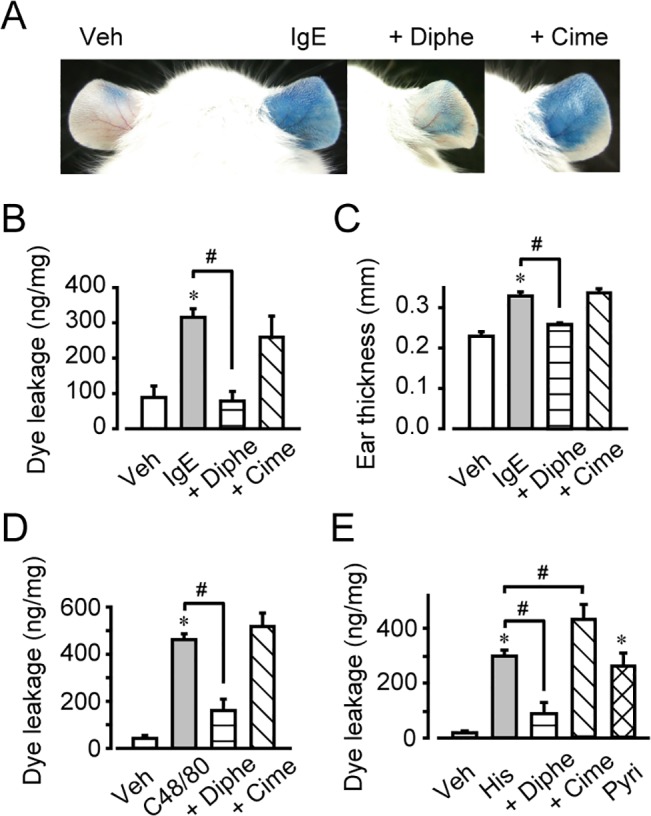
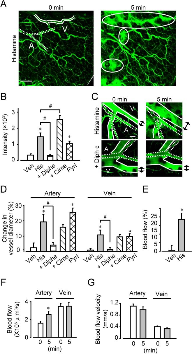
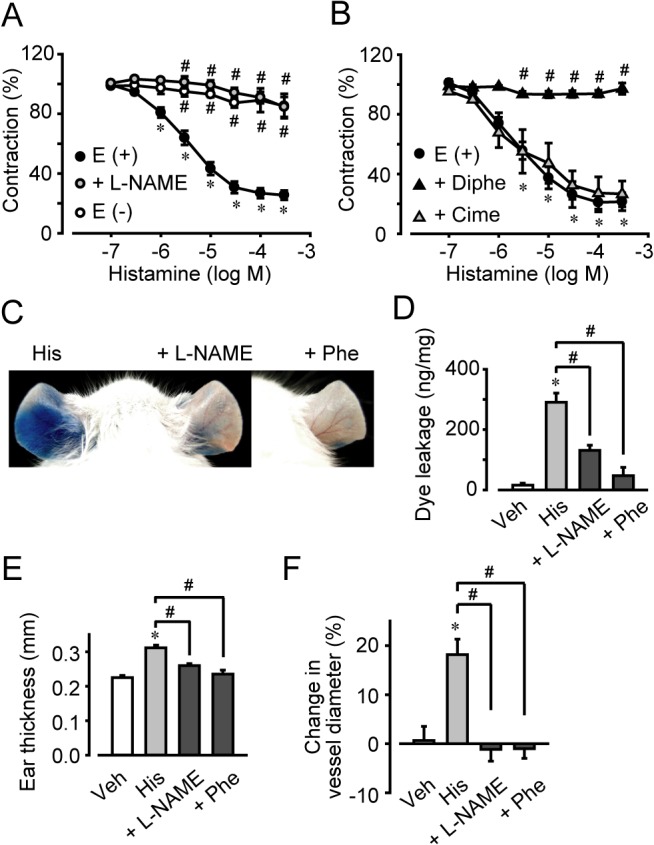
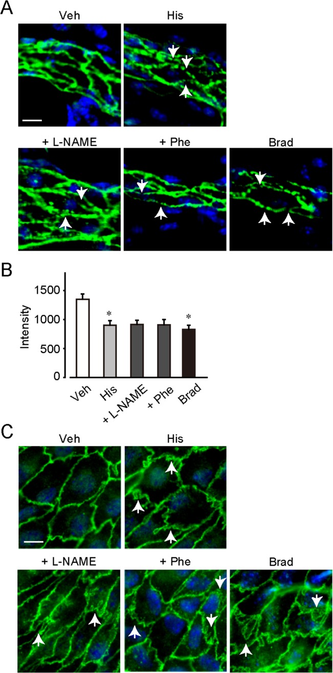
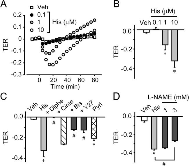
Histamine is a mediator of allergic inflammation released mainly from mast cells. Although histamine strongly increases vascular permeability, its precise mechanism under in vivo situation remains unknown. We here attempted to reveal how histamine induces vascular hyperpermeability focusing on the key regulators of vascular permeability, blood flow and endothelial barrier. Degranulation of mast cells by antigen-stimulation or histamine treatment induced vascular hyperpermeability and tissue swelling in mouse ears. These were abolished by histamine H1 receptor antagonism. Intravital imaging showed that histamine dilated vasculature, increased blood flow, while it induced hyperpermeability in venula. Whole-mount staining showed that histamine disrupted endothelial barrier formation of venula indicated by changes in vascular endothelial cadherin (VE-cadherin) localization at endothelial cell junction. Inhibition of nitric oxide synthesis (NOS) by L-NAME or vasoconstriction by phenylephrine strongly inhibited the histamine-induced blood flow increase and hyperpermeability without changing the VE-cadherin localization. In vitro, measurements of trans-endothelial electrical resistance of human dermal microvascular endothelial cells (HDMECs) showed that histamine disrupted endothelial barrier. Inhibition of protein kinase C (PKC) or Rho-associated protein kinase (ROCK), NOS attenuated the histamine-induced barrier disruption. These observations suggested that histamine increases vascular permeability mainly by nitric oxide (NO)-dependent vascular dilation and subsequent blood flow increase and maybe partially by PKC/ROCK/NO-dependent endothelial barrier disruption.


Conflict of interest statement

Competing Interests: The authors have declared that no competing interests exist.

Figures

Fig 1
Fig 1. H1 receptor activation increased vascular permeability.
The effects of diphenhydramine (Diphe) or cimetidine (Cime), 2-pyridylethylamine (Pyri) on vascular hyperpermeability. (A) Photographs of representative mouse ears. (B) Quantification of Evans blue leakage (n = 4–6). (C) Quantification of ear thickness (n = 4–6). #P < 0.05, as compared with IgE. (D) Quantification of Evans blue leakage after compound 48/80 (C48/80) application (n = 4–6). (E) Quantification of Evans blue leakage after histamine application (n = 4–7). *P < 0.05, compared with vehicle. #P < 0.05, compared with histamine. Data are presented as mean ± SEM.
Fig 2
Fig 2. Two types of vasculature are in mouse ear.
(A) Observation point. (B) Whole-mount immunostaining of PECAM-1, α-SMA, and desmin in the proximal vessel (magnification, ×200). Bar, 100 μm. A, artery; V, vein. (C) Whole-mount immunostaining of PECAM-1, α-SMA, desmin, and FCεRI in the capillary (magnification, ×200). Bar, 100 μm. Dotted lines indicate the edge of ear.
Fig 3
Fig 3. Histamine increased blood flow volume.
(A) Typical images of the proximal vessel region before and 5 min after histamine treatment (magnification, ×100). Bar, 200 μm. Solid lines indicate vein. Dotted lines indicate artery. Ovoid circles indicate the FITC-dextran leakage. (B) Quantification of the FITC-dextran leakage after histamine application (n = 6–12). (C) Typical images of histamine-induced relaxation (magnification, ×200). Bar, 100 μm. A, artery; V, vein. (D) Quantification of the change in vessel diameter (n = 6–12). (E) Measurement of blood flow 5 min after histamine application using laser doppler velocimetry (n = 8). Measurement of blood flow (F) and blood flow velocity (G) before and 5 min after histamine application using in vivo microscopy (n = 15). *P < 0.05, compared with vehicle. #P < 0.05, compared with histamine. Data are presented as mean ± SEM.
Fig 4
Fig 4. L-NAME or phenylephrine pretreatment inhibited histamine-induced hyperpermeability and vascular relaxation.
(A) Effect of L-NAME pretreatment or endothelium on the histamine-induced relaxation of mesenteric artery. (B) Effect of diphenhydramine or cimetidine pretreatment on the histamine-induced relaxation of mesenteric artery(n = 4–5). (C) Effect of L-NAME or phenylephrine (Phe) on histamine-induced vascular hyperpermeability. Typical photographs showing extravasation of Evans blue after histamine treatment. (D) Quantification of the Evans blue leakage (n = 4–7). (E) Quantification of the ear thickness (n = 4–6). (F) Quantification of arterial diameter changes (n = 6–12). *P < 0.05, compared with vehicle. #P < 0.05, compared with histamine. Data are presented as mean ± SEM.
Fig 5
Fig 5. Histamine regulated endothelial barrier function in vivo.
(A) Whole-mount immunostaining of VE cadherin in the ear vessel (magnification, ×400). Bar, 10 μm. (B) Fluorescence intensity of VE-cadherin at endothelial cell junction in ear vessel (n = 4–5). (C) En face immunostaining of VE-cadherin in the pulmonary artery (magnification, ×400). Bar, 10 μm. *P < 0.05, compared with vehicle. Data are presented as mean ± SEM.
Fig 6
Fig 6. Histamine regulated endothelial barrier function in vitro.
(A) Typical graph of histamine treatment in HDMECs. (B) Quantification of TER in HDMECs (n = 4–8). (C) Effects of pretreatment with diphenhydramine, cimetidine, Y27632, and bisindolylmaleimide 1 on the histamine-induced change in transendothelial electric resistance (TER) in HDMECs (n = 4–6). (D) Effect of pretreatment with L-NAME on the histamine-induced change in TER in HDMECs (n = 4). *P < 0.05, compared with non-treated control. #P < 0.05, compared with histamine. Data are presented as mean ± SEM.

References

    1. Curry FR, Adamson RH. Vascular permeability modulation at the cell, microvessel, or whole organ level: towards closing gaps in our knowledge. Cardiovasc Res. 2010;87(2):218–29. Epub 2010/04/27. 10.1093/cvr/cvq115 - DOI - PMC - PubMed
    1. Nathan RA. The pathophysiology, clinical impact, and management of nasal congestion in allergic rhinitis. Clin Ther. 2008;30(4):573–86. Epub 2008/05/24. . - PubMed
    1. Feletou M, Bonnardel E, Canet E. Bradykinin and changes in microvascular permeability in the hamster cheek pouch: role of nitric oxide. Br J Pharmacol. 1996;118(6):1371–6. Epub 1996/07/01. - PMC - PubMed
    1. Wallez Y, Huber P. Endothelial adherens and tight junctions in vascular homeostasis, inflammation and angiogenesis. Biochimica et biophysica acta. 2008;1778(3):794–809. Epub 2007/10/27. 10.1016/j.bbamem.2007.09.003 . - DOI - PubMed
    1. van Nieuw Amerongen GP, van Delft S, Vermeer MA, Collard JG, van Hinsbergh VW. Activation of RhoA by thrombin in endothelial hyperpermeability: role of Rho kinase and protein tyrosine kinases. Circulation research. 2000;87(4):335–40. Epub 2000/08/19. . - PubMed

Publication types

MeSH terms