. 2015 Jul 28;112(30):9436-41.
doi: 10.1073/pnas.1508686112. Epub 2015 Jul 9.

Structural basis and functional analysis of the SARS coronavirus nsp14-nsp10 complex

Affiliations

Structural basis and functional analysis of the SARS coronavirus nsp14-nsp10 complex

Yuanyuan Ma et al. Proc Natl Acad Sci U S A. .

Abstract

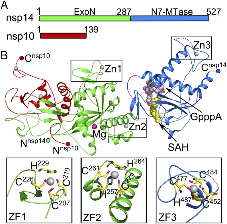
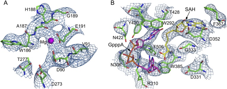
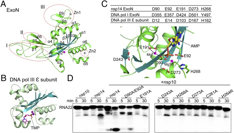
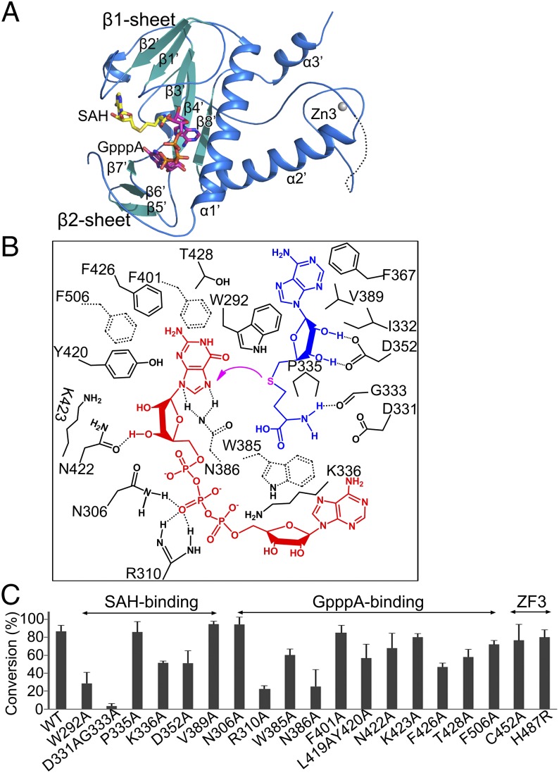
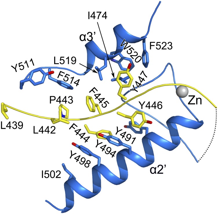
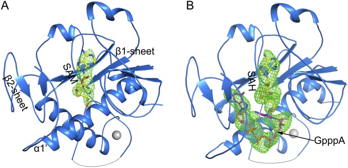
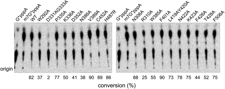
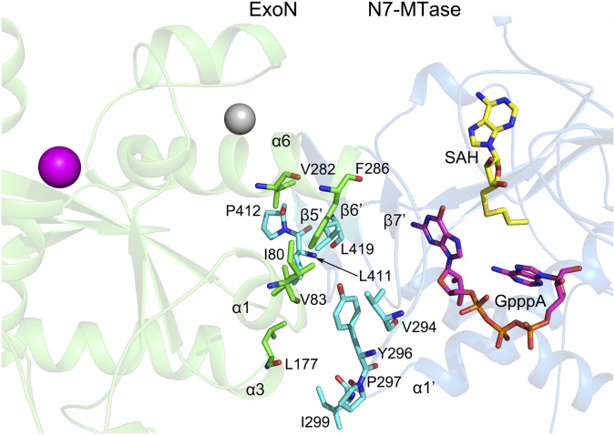
Nonstructural protein 14 (nsp14) of coronaviruses (CoV) is important for viral replication and transcription. The N-terminal exoribonuclease (ExoN) domain plays a proofreading role for prevention of lethal mutagenesis, and the C-terminal domain functions as a (guanine-N7) methyl transferase (N7-MTase) for mRNA capping. The molecular basis of both these functions is unknown. Here, we describe crystal structures of severe acute respiratory syndrome (SARS)-CoV nsp14 in complex with its activator nonstructural protein10 (nsp10) and functional ligands. One molecule of nsp10 interacts with ExoN of nsp14 to stabilize it and stimulate its activity. Although the catalytic core of nsp14 ExoN is reminiscent of proofreading exonucleases, the presence of two zinc fingers sets it apart from homologs. Mutagenesis studies indicate that both these zinc fingers are essential for the function of nsp14. We show that a DEEDh (the five catalytic amino acids) motif drives nucleotide excision. The N7-MTase domain exhibits a noncanonical MTase fold with a rare β-sheet insertion and a peripheral zinc finger. The cap-precursor guanosine-P3-adenosine-5',5'-triphosphate and S-adenosyl methionine bind in proximity in a highly constricted pocket between two β-sheets to accomplish methyl transfer. Our studies provide the first glimpses, to our knowledge, into the architecture of the nsp14-nsp10 complex involved in RNA viral proofreading.

Keywords: CoV; exoribonuclease; methyltransferase; nsp14; proofreading.


Conflict of interest statement

The authors declare no conflict of interest.

Figures

Fig. 1.
Fig. 1.
Overall structure of the nsp14–nsp10 complex. (A) Domain organization of nsp14 and nsp10. Domain boundaries are marked with residue numbers. (B) Cartoon representation of the structure of the nsp14–nsp10 heterodimer. Nsp10, the ExoN domain, the N7-MTase domain, and the loop at the N terminus of N7-MTase are marked red, green, marine, and pink, respectively. Invisible residues from 454 to 464 of nsp14 are shown by a dashed line. Magnesium, zinc ions, and the N7-MTase substrates SAH and GpppA are shown as spheres and are colored magenta, gray, yellow, and light pink, respectively. Three zinc fingers (ZF) of nsp14 are highlighted with residues shown as sticks.
Fig. S1.
Fig. S1.
Purification of the nsp14–nsp10 complex. During coexpression in E. coli, nsp10 was expressed in excess of nsp14. The nsp14–nsp10 complex was separated from the excess nsp10 by HiTrap S ion-exchange chromatography. Nsp10 did not bind to the column (Peak 1). Peak 2 eluted from the column using NaCl contained the complex of nsp14 with nsp10. (Inset) SDS/PAGE.
Fig. S2.
Fig. S2.
2Fo-Fc maps of the active centers of nsp14. The electron density for the ExoN catalytic center (A) and the N7-MTase catalytic center (B) are contoured at 2σ. Amino acids surrounding the catalytic centers and substrates SAH and GpppA are shown as sticks, and Mg2+ is shown as a magenta sphere.
Fig. 2.
Fig. 2.
Comparison of the structure and catalytic residues of nsp14 ExoN domain with proofreading homologs. (A) Cartoon representation of the ExoN domain marked with secondary structural elements. The three different regions from other DEDD superfamily exonucleases are indicated by red dashed ellipses. (B) The structure of the E. coli ε subunit of polymerase III (Pol III) is shown in the same orientation as nsp14 for comparison. Metal ions are shown as spheres, and bound ligands are shown as sticks. (C) The active center of the ExoN domain of nsp14. (Upper) Catalytic residues of the ExoN domain of nsp14, the exonuclease domain of DNA polymerase I, and the ε subunit of DNA polymerase III of E. coli are listed in the table. (Lower) Catalytic residues, the modeled substrate AMP, and the mistaken D243 are shown as sticks. MgB observed in the structure and MgA modeled are shown as spheres. Dashed lines indicate the hydrogen bonds between MgB and D90 and E191. (D) Exoribonuclease assays for nsp10, nsp14 alone, and nsp14 or nsp14 mutants in complex with nsp10 on 5′-labeled ssRNA of 22 nucleosides (RNA22). The symbol “#” indicates cleavage products.
Fig. S3.
Fig. S3.
Primary sequence alignment of nsp14 homologs by ClustalW (53) and rendered using ESPript version 3.0 (54). The secondary structural elements and numbering of SARS-CoV nsp14 are displayed above the alignment. Identical residues in all sequences are boxed with red background; those conserved are marked in red. Catalytic residues, D243, and residues involved in formation of zinc fingers are marked with magenta stars, red stars, and green triangles, respectively. Sequences of nsp14 in SARS-CoV (accession number ADC35510), MERS-CoV (AIZ48758), mouse hepatitis virus (MHV; NP_068668), human coronavirus [(HCoV)-229E; (AGT21366.1], and infectious bronchitis virus (IBV; P0C6Y2) were used for the analysis.
Fig. 3.
Fig. 3.
Intermolecular interactions between nsp14 and nsp10. Zinc ions are represented as gray spheres in AC. (A) The Nsp14 ExoN domain (green) is stabilized by nsp10 (red). Two regions of nsp10 (boxed) contribute major interactions with nsp14. Zn1nsp10, the first zinc ion of nsp10. (B and C) Interaction details by regions 1 (B) and 2 (C). Hydrogen bonds between residues are shown by dashed lines. Residues of nsp14 and nsp10 involved in interaction are displayed as green and red sticks, respectively. (D) Schematic representation of the contacts between nsp10 and nsp14.
Fig. S4.
Fig. S4.
Comparison of the nsp16 and nsp14 interaction sites on the nsp10 surface. Nsp10 of the nsp14–nsp10–SAM complex is shown as a gray surface and cartoon. The interaction sites for nsp10 with nsp16 and nsp14 are colored in salmon and orange, respectively.
Fig. 4.
Fig. 4.
Structure and methyl transfer mechanism of the nsp14 N7-MTase domain. (A) Cartoon representation of the N7-MTase domain marked with secondary structural elements. (B) Amino acids within 4 Å of the ligands SAH (blue) and GpppA (red) are labeled and numbered. The magenta arrow indicates the methyl transfer. Dashed lines between residues indicate hydrogen bonds. Trp385, Asn386, Phe401, and Phe506 are shown by dashed bonds to depict their position below the plane of ligand GpppA. (C) The ability of nsp14 to methylate N7 of guanine of GpppA-RNA was measured. The results depict the efficiency of the conversion of substrate to product (%) and are plotted as a bar graph. WT nsp14 and its mutants were complexed with nsp10.
Fig. S5.
Fig. S5.
C-terminal hydrophobic interaction network of the N7-MTase domain of nsp14. The less stable region between Leu439 and Glu453 (shown in yellow) interacts with α2′ and the last α-helix α3′ via hydrophobic interactions. Key residues involved in formation of the hydrophobic network are shown as sticks and are labeled.
Fig. S6.
Fig. S6.
Ligand-binding sites of the nsp14 N7-MTase domain. Substrates SAM (A) and GpppA and SAH (B) bind in pockets formed by the β1-sheet, β2-sheet, and α1′. Fo-Fc electron density maps (omit map) for SAM, GpppA, and SAH contoured at 2σ are shown.
Fig. S7.
Fig. S7.
N7-MTase activity of nsp14 and its mutants. Nsp14 and its mutants were tested for their ability to methylate 32P-labeled G*pppA-RNA. After incubation for 6 min at 37 °C, the reaction products were digested by nuclease P1 into single nucleotides and methylated m7G*pppA or unmethylated G*pppA. They were analyzed by separating the mixture using TLC. The position of origin is marked on the left. The percentage of conversion of G*pppA-RNA to m7G*pppA-RNA is listed at the bottom.
Fig. S8.
Fig. S8.
The hydrophobic interactions between the ExoN domain and N7-MTase domain. Interfacing residues between the ExoN and N7-MTase domains are displayed as green and cyan sticks, respectively. Secondary structural elements involved in the interactions are labeled.
Fig. 5.
Fig. 5.
Model for the role of nsp14 in proofreading and mRNA capping. Nascent RNA is synthesized at the polymerization sites (red circle) of the nsp12 RdRp domain (gray) or is mismatch excised at the proofreading site (red circles) of nsp14 ExoN (green surface). The relative orientation of the polymerization and proofreading domains is built based on the Klenow fragment. The 5′ end of newly synthesized mRNA is modified by sequential activities contributed by RTPase of nsp13, GTase (currently unknown), N7-MTase of nsp14, and 2′-O–MTase of nsp16 (light pink surface, PDB ID code 3R24) for formation of a cap1 structure.

References

    1. De Groot R, et al. Order nidovirales. In: King AM, Adams MJ, Lefkowitz EJ, editors. Virus Taxonomy, Ninth Report of the International Committee on Taxonomy of Viruses. Elsevier; London: 2011. pp. 785–795.
    1. Peiris JS, et al. SARS study group Coronavirus as a possible cause of severe acute respiratory syndrome. Lancet. 2003;361(9366):1319–1325. - PMC - PubMed
    1. Zaki AM, van Boheemen S, Bestebroer TM, Osterhaus AD, Fouchier RA. Isolation of a novel coronavirus from a man with pneumonia in Saudi Arabia. N Engl J Med. 2012;367(19):1814–1820. - PubMed
    1. Gorbalenya AE, Enjuanes L, Ziebuhr J, Snijder EJ. Nidovirales: Evolving the largest RNA virus genome. Virus Res. 2006;117(1):17–37. - PMC - PubMed
    1. Snijder EJ, et al. Unique and conserved features of genome and proteome of SARS-coronavirus, an early split-off from the coronavirus group 2 lineage. J Mol Biol. 2003;331(5):991–1004. - PMC - PubMed

Publication types

MeSH terms

LinkOut - more resources