

Histologic Analysis of Retrieved Clots in Acute Ischemic Stroke: Correlation with Stroke Etiology and Gradient-Echo MRI
- PMID: 26159515
- PMCID: PMC7968760
- DOI: 10.3174/ajnr.A4402
Histologic Analysis of Retrieved Clots in Acute Ischemic Stroke: Correlation with Stroke Etiology and Gradient-Echo MRI
Abstract
Background and purpose: It is unclear whether clot composition analysis is helpful to predict a stroke mechanism in acute large vessel occlusion. In addition, the relationship between early vessel signs on imaging studies and clot compositions has been poorly understood. The purpose of this study was to elucidate the relationship between clot composition and stroke etiology following mechanical thrombectomy and to investigate the effect of varied clot compositions on gradient-echo MR imaging of clots.
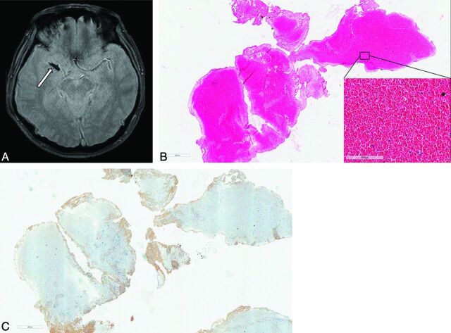
Materials and methods: Histopathologic analysis of retrieved clots from 37 patients with acute MCA occlusion was performed. Patients underwent gradient-echo imaging before endovascular therapy. Retrieved clots underwent semiquantitative proportion analysis to quantify red blood cells, fibrin, platelets, and white blood cells by area. Correlations between clot compositions and stroke subtypes and susceptibility vessel signs on gradient-echo imaging were assessed.
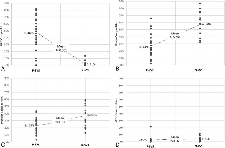
Results: Stroke etiology was classified as cardioembolism in 22 patients (59.4%), large-artery atherosclerosis in 8 (21.6%), and undetermined in 7 (18.9%). The clots from cardioembolism had a significantly higher proportion of red blood cells (37.8% versus 16.9%, P = .031) and a lower proportion of fibrin (32.3% versus 48.5%, P = .044) compared with those from large-artery atherosclerosis. The proportion of red blood cells was significantly higher in clots with a susceptibility vessel sign than in those without it (48.0% versus 1.9%, P < .001), whereas the proportions of fibrin (26.4% versus 57.0%, P < .001) and platelets (22.6% versus 36.9%, P = .011) were significantly higher in clots without a susceptibility vessel sign than those with it.
Conclusions: The histologic composition of clots retrieved from cerebral arteries in patients with acute stroke differs between those with cardioembolism and large-artery atherosclerosis. In addition, a susceptibility vessel sign on gradient-echo imaging is strongly associated with a high proportion of red blood cells and a low proportion of fibrin and platelets in retrieved clots.
© 2015 by American Journal of Neuroradiology.
Figures


References
-
- Saver JL, Jahan R, Levy EI, et al. ; SWIFT Trialists. Solitaire flow restoration device versus the Merci retriever in patients with acute ischaemic stroke (SWIFT): a randomised, parallel-group, non-inferiority trial. Lancet 2012;380:1241–49 - PubMed
-
- Turk AS, Frei D, Fiorella D, et al. . ADAPT FAST study: a direct aspiration first pass technique for acute stroke thrombectomy. J Neurointerv Surg 2014;6:260–64 - PubMed
-
- Kim SK, Yoon W, Moon SM, et al. . Outcomes of manual aspiration thrombectomy for acute ischemic stroke refractory to stent-based thrombectomy. J Neurointerv Surg 2015;7:473–77 - PubMed
MeSH terms
Substances
LinkOut - more resources
Full Text Sources
Medical