. 2015 Jul 8;18(1):27-37.
doi: 10.1016/j.chom.2015.06.011.

Innate Immune Defenses Mediated by Two ILC Subsets Are Critical for Protection against Acute Clostridium difficile Infection

Affiliations

Innate Immune Defenses Mediated by Two ILC Subsets Are Critical for Protection against Acute Clostridium difficile Infection

Michael C Abt et al. Cell Host Microbe. .

Abstract

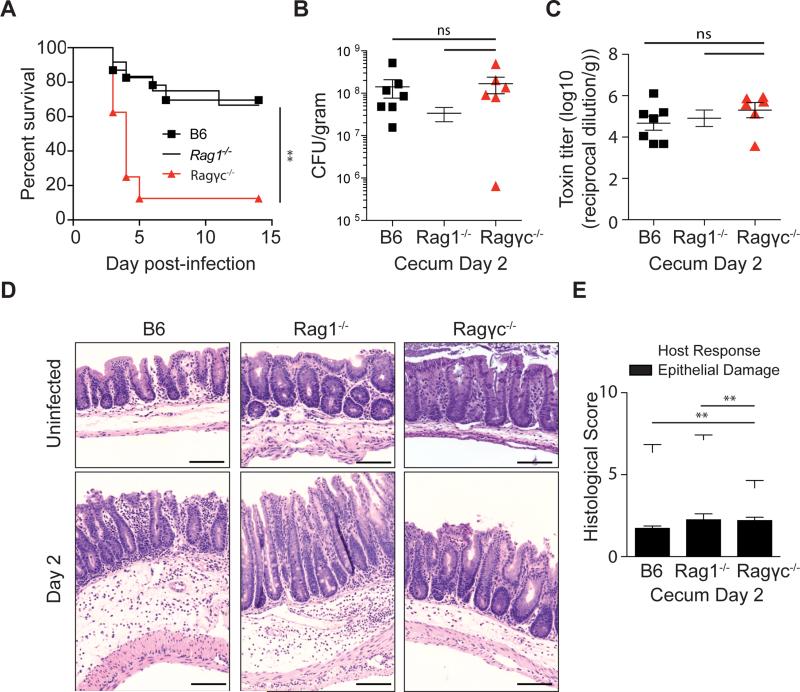
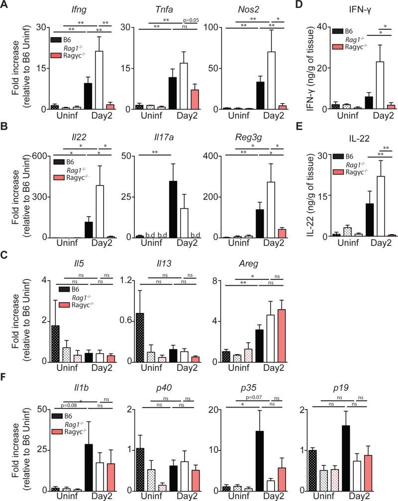
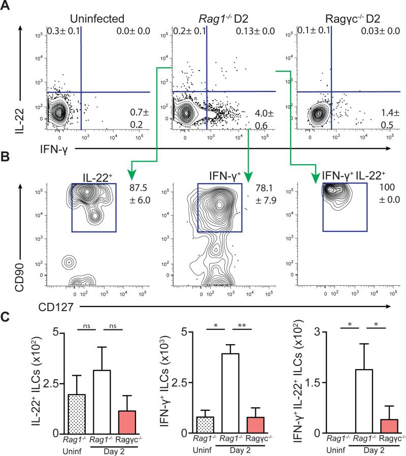
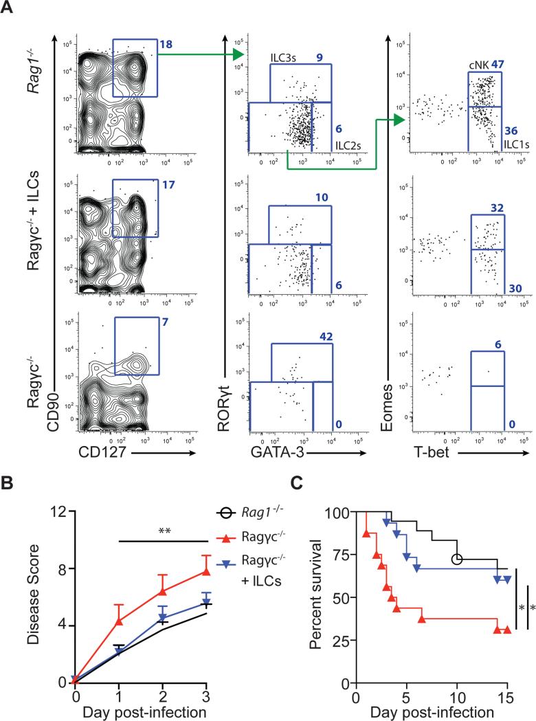
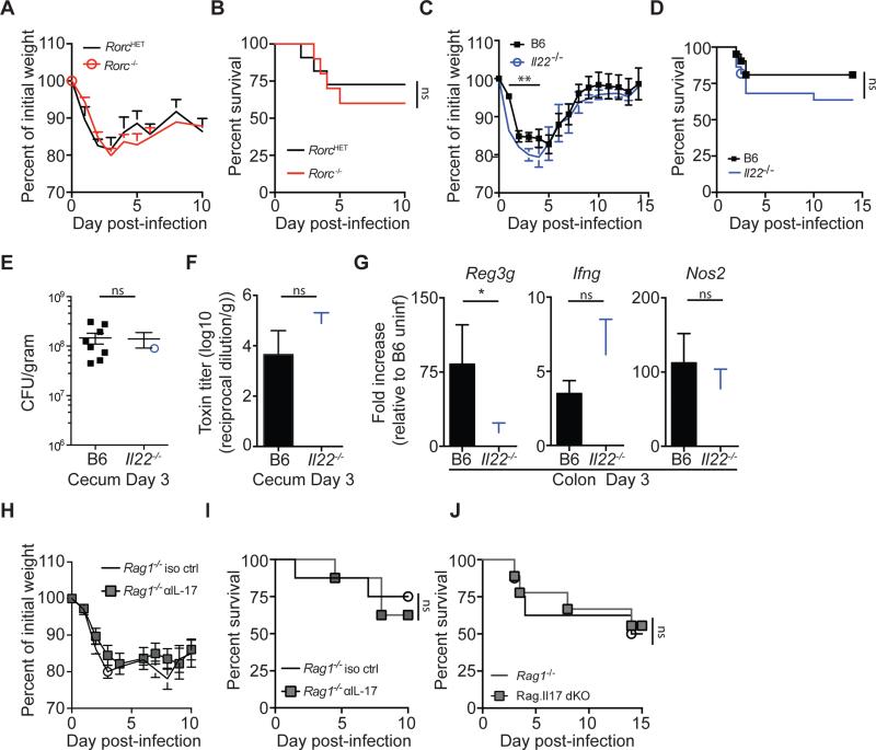
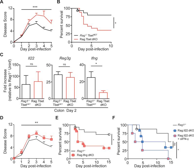
Infection with the opportunistic enteric pathogen Clostridium difficile is an increasingly common clinical complication that follows antibiotic treatment-induced gut microbiota perturbation. Innate lymphoid cells (ILCs) are early responders to enteric pathogens; however, their role during C. difficile infection is undefined. To identify immune pathways that mediate recovery from C. difficile infection, we challenged C57BL/6, Rag1(-/-) (which lack T and B cells), and Rag2(-/-)Il2rg(-/-) (Ragγc(-/-)) mice (which additionally lack ILCs) with C. difficile. In contrast to Rag1(-/-) mice, ILC-deficient Ragγc(-/-) mice rapidly succumbed to infection. Rag1(-/-) but not Ragγc(-/-) mice upregulate expression of ILC1- or ILC3-associated proteins following C. difficile infection. Protection against infection was restored by transferring ILCs into Ragγc(-/-) mice. While ILC3s made a minor contribution to resistance, loss of IFN-γ or T-bet-expressing ILC1s in Rag1(-/-) mice increased susceptibility to C. difficile. These data demonstrate a critical role for ILC1s in defense against C. difficile.


Figures

Figure 1
Figure 1. Absence of innate lymphoid cells leads to increased susceptibility to C. difficile
(A) C57BL/6, Rag1−/−, Ragγc−/− mice were inoculated with 200 spores of C. difficile (VPI 10463 strain) and were assessed for survival following infection. Survival curve is a combination of 5 independent experiments (C57BL/6 n=23, Rag1−/− n=12, Ragγc−/− n=8). At day 2 post-infection (B) C. difficile burden and (C) toxin levels were measured from the cecal content. (D) Representative H&E stained cecal sections from antibiotic-treated, uninfected and day 2 infected C57BL/6, Rag1−/−, Ragγc−/− mice. Scale bar = 100μm. (E) Pathology score of histological tissue sections based on cellular infiltration/edema (host response), and epithelial layer degeneration (n=9-11). *p<0.05, **p<0.01. ns = not significant. Data shown are mean ± SEM. See also Figure S1 and S2.
Figure 2
Figure 2. C57BL/6 and Rag1−/−, but not Ragγc−/− mice upregulate expression of ILC1 or ILC3 associated proteins following C. difficile infection
Fold Induction of (A) ILC1, (B) ILC3, (C) ILC2 associated effector molecules in the colon at day 2 post-infection relative to uninfected C57BL/6 mice and normalized to Hprt. (D) IFN-γ and (E) IL-22 protein in the supernatant of cecal tissue explants. (F) Fold induction of genes upstream of ILC1 and ILC3 activation (n=7-13) *p<0.05, **p<0.01. Data shown are mean ± SEM. b.d. = below detection.
Figure 3
Figure 3. IL-22 and IFN-γ expressing ILCs are induced in the large intestine following C. difficile infection
Cells isolated from the large intestine intraepithelial compartment of uninfected Rag1−/−, day 2 infected Rag1−/−, or day 2 infected Ragγc−/− mice were incubated in media in the presence of brefeldin A (BFA) and assessed for IL-22 and IFN-γ production. (A) Frequency of IL-22+, IFN-γ+, and IL-22+/ IFN-γ+ cells. FACS plots gated on live, CD45+, Gr-1neg cells. (B) Expression of CD90 and CD127 on cytokine positive cells from day 2 infected Rag1−/− mice. (C) Number of IL-22+, IFN-γ+, and IL-22+/IFN-γ+ ILCs. Data representative of 3 independent experiments (n=4-5.) *p<0.05, **p<0.01. Data shown are mean ± SEM. See also Figure S3.
Figure 4
Figure 4. Adoptive transfer of ILCs into Ragγc−/− mice restores host defense following C. difficile infection
(A) Reconstitution of ILC compartment in the large intestine Lp of recipient Ragγc−/− mice 3 weeks after ILC transfer. Rag1−/−, Ragγc−/−, and Ragγc−/− +ILCs mice were infected with C. difficile and were assessed for (B) disease severity and (C) survival following infection. Combination of 5 independent experiments (n=15-18). *p<0.05, **p<0.01. Data shown are mean ± SEM. See also Figure S4.
Figure 5
Figure 5. Selective loss of ILC3s or ILC3 effector molecules reveals their limited role in recovery following acute C. difficile infection
(A) Weight loss and (B) survival of RorcHET or Rorc KO mice following CDI. Combination of 3 independent experiments (n=10-11). (C-G) C57BL/6 and Il22−/− mice were infected with C. difficile spores and assessed for (C) weight loss and (D) survival. Weight loss curve is representative of 4 independent experiments. Survival curve is a combination of 4 experiments (n=21-22). (E) C. difficile burden and (F) toxin levels in the cecal content at day 3 post-infection. (G) Fold induction of Reg3g, Ifng, and Nos2 in the colon of day 3 infected C57BL/6 and Il22−/− mice relative to uninfected C57BL/6 mice and normalized to Hprt (n=7-8). (H) Weight loss and (I) survival of Rag1−/− or Rag1−/− mice treated with anti-IL-17a neutralizing antibody (n=8). (J) Survival of Rag1−/− or Rag.Il17 dKO mice (n=7-9). *p<0.05, **p<0.01. Data shown are mean ± SEM. See also Figure S5.
Figure 6
Figure 6. Ablation of ILC1s or ILC1-associated IFN-γ leads to increased susceptibility to C. difficile infection
(A) Disease severity and (B) survival of Rag1−/− TbetHET or Rag.Tbet dKO mice following CDI. Combination of 3 independent experiments (Rag1−/− TbetHET n=14, Rag.Tbet dKO n=16). (C) Fold induction of Il22, Reg3g, and Ifng in the colon of day 2 infected Rag1−/− TbetHET or Rag.Tbet dKO mice relative to uninfected Rag1−/− TbetHET mice and normalized to Hprt (n=7). (D) Disease severity and (E) survival of Rag1−/− or Rag.Ifng dKO mice following CDI. Combination of 4 independent experiments (n=15-17). (F) Survival of Rag1−/−, Rag.Il22 dKO or Rag.Il22 dKO mice treated with αIFN-γ neutralizing antibody (n=4-8). *p<0.05, **p<0.01, ***p<0.001. Data shown are mean ± SEM. See also Figure S6.

Comment in

References

    1. Artis D, Spits H. The biology of innate lymphoid cells. Nature. 2015;517:293–301. - PubMed
    1. Bernink JH, Peters CP, Munneke M, te Velde AA, Meijer SL, Weijer K, Hreggvidsdottir HS, Heinsbroek SE, Legrand N, Buskens CJ, et al. Human type 1 innate lymphoid cells accumulate in inflamed mucosal tissues. Nature immunology. 2013;14:221–229. - PubMed
    1. Boyman O, Ramsey C, Kim DM, Sprent J, Surh CD. IL-7/anti-IL-7 mAb complexes restore T cell development and induce homeostatic T Cell expansion without lymphopenia. Journal of immunology. 2008;180:7265–7275. - PubMed
    1. Buffie CG, Bucci V, Stein RR, McKenney PT, Ling L, Gobourne A, No D, Liu H, Kinnebrew M, Viale A, et al. Precision microbiome reconstitution restores bile acid mediated resistance to Clostridium difficile. Nature. 2015;517:205–208. - PMC - PubMed
    1. Buffie CG, Jarchum I, Equinda M, Lipuma L, Gobourne A, Viale A, Ubeda C, Xavier J, Pamer EG. Profound alterations of intestinal microbiota following a single dose of clindamycin results in sustained susceptibility to Clostridium difficile-induced colitis. Infection and immunity. 2012;80:62–73. - PMC - PubMed

Publication types

LinkOut - more resources