. 2015 Sep;60(9):1416-27.
doi: 10.1016/j.archoralbio.2015.06.007. Epub 2015 Jun 20.

Decrease of miR-146a is associated with the aggressiveness of human oral squamous cell carcinoma

Affiliations

Decrease of miR-146a is associated with the aggressiveness of human oral squamous cell carcinoma

Zonggao Shi et al. Arch Oral Biol. 2015 Sep.

Abstract

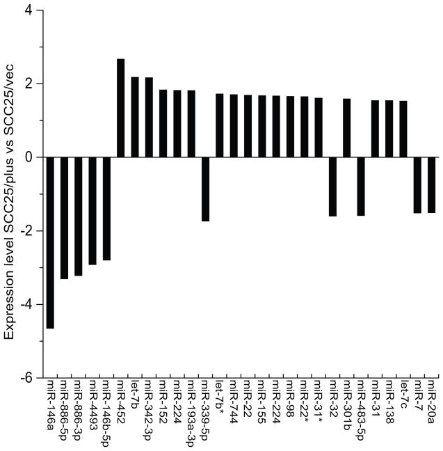
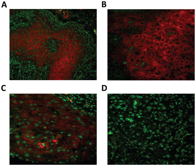
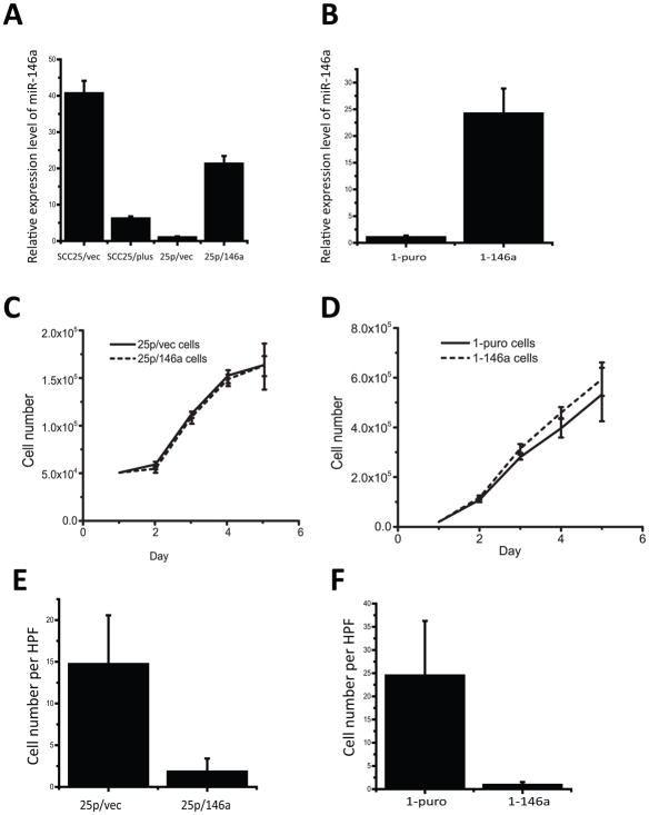
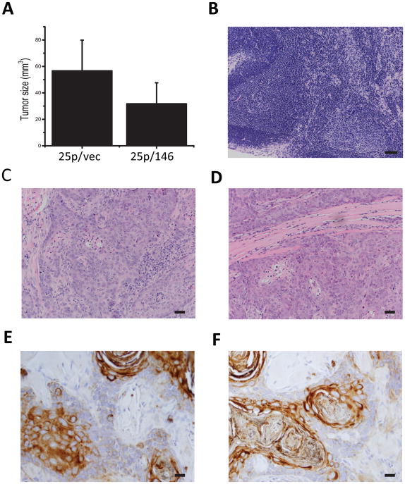
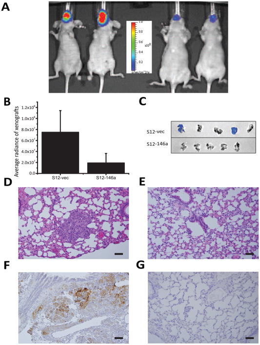
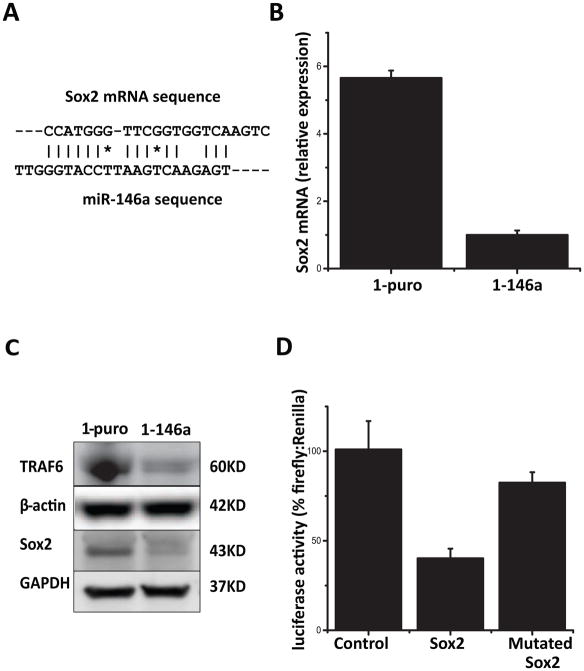
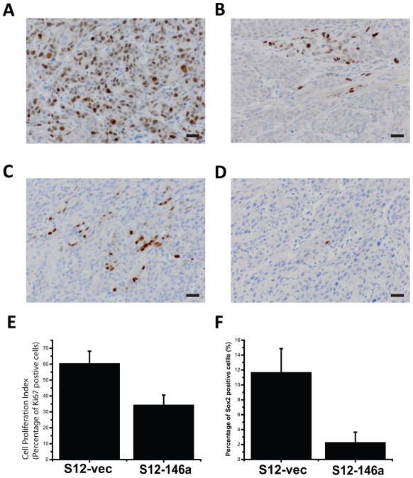
With the aim to identify microRNAs that may contribute to oral squamous cell carcinoma (OSCC) progression, we compared the microRNA expression profiles of two related cell lines that form tumors with differential aggressiveness. A panel of 28 microRNAs was found to be more than 1.5-fold altered, among which miR-146a was the most significantly changed (-4.6-fold). Loss of miR-146a expression was validated in human high-grade tumors, while normal oral mucosa retained expression, using fluorescence in situ hybridization on a tissue microarray. Restoration of miR-146a in SCC25 and UMSCC1 cells decreased in vitro invasive activity, suppressed tumor growth in vivo, and decreased the incidence of UMSCC1 lung metastasis. The transcription factor Sox2 was found to be a putative target of miR-146a. In conclusion, the loss or decrease of miR-146a is a new feature that is associated with more aggressive behaviour in oral squamous carcinoma.

Keywords: Metastasis; MicroRNA; Oral cancer; miR-146a.


Figures

Figure 1
Figure 1. Comparative microRNA profile of oral cancer cell lines
Expression profiles of SCC25/plus cells, which overexpress PLAUR and grow as invasive poorly differentiated tumors in vivo were compared to parental SCC25 cells (designated SCC25/vec). Total RNA samples were extracted from SCC25/vec and SCC25/plus cells and microRNA was performed in duplicate on Exiqon’s miRCURY LNA microRNA arrays (v.10.0). Expression level is presented as the log 2 ratio of SCC25/plus vs SCC25/vec cells.
Figure 2
Figure 2. Fluorescence in situ hybridization (FISH) detection of miR-146a in human OSCC
Tissue microarray sections containing 10 human normal oral mucosa cores and 50 oral squamous carcinoma (OSCC) cores were hybridized with LNA-based digoxigenin labeled miR-146a probe. Positive signal was amplified by tyramide signal amplification system (Cy5) and visualized with fluorescence microscopy. Shown are photomicrographs from (A) normal squamous epithelium, (B) OSCC histologic grade 1, (C) OSCC grade 2 and (D) OSCC grade 3. Magnification 400×, scale bar=20μm.
Figure 3
Figure 3. Restoration of miR-146a expression inhibits oral cancer cell invasion
(A) Quantitative PCR (pPCR) analysis of miR-146a expression validates loss of miR-146a in SCC25/plus relative to parental SCC25/vec cells. Using viral vectors, expression of miR-146a was restored to physiologic levels in SCC25/plus cells (designated 25p/146a), relative to vector controls (25p/vec). (B) Viral vectors were used to restore miR-146a expression in UMSCC1 cells (designated 1-146a) relative to vector controls (1-puro). Levels of miR-146a were assessed by qPCR. (*) p<0.05. (C), (D) Growth curves of (C) 25p/vec, 25p/146a and (D) 1-puro, 1-146a cells were obtained by daily counting. No statistically significant differences in proliferation were observed. (E), (F) Boyden chamber Matrigel invasion assays for cells derived from (E) SCC25/plus (25p/vec vs 25/146a) and (F) UMSCC1 cells (1-puro vs 1-146a). 250,000 cells in 500μl per well were added to the upper chamber and incubated for 24 h prior to stopping the assay using Diff-quick stain and enumeration of cells migrating to the lower chamber. The average cell numbers per high-power field (HPF) were compared. A significant decrease (p<.05) in invasion is observed in cells expressing miR-146a.
Figure 4
Figure 4. Restoration of miR-146a inhibits tumorigenicity in SCC25 xenografts
Using an orthotopic xenograft model, the tumor forming ability of SCC25 derived 25p/vec and 25p/146a cells was tested by tongue injection. Cells (1.2×10⁶ cells in 30μl) were injected and tumor growth was monitored for 8 weeks prior to sacrifice and analysis. (A) Tumor volume (V) was calculated based on measured longest diameter (a) and the perpendicular (b) with calipers using formula: V=(a*b^2)/2. A significant difference in tumor volume was observed between tumors generated from 25p/vec and 25p/146a cells, P<0.05. (B) Hematoxylin & eosin (H&E) staining of a representative lymph node. Magnification 200×, scale bar=50μm. (C, D) H&E staining of primary tumor generated by (C) 25p/vec and (D) 25p/146a cells. Magnification 200×. (E, F) Immunohistochemical staining of pan-cytokeratin (AE1/AE3) on tumors derived from (E) 25p/vec and (F) 25p/146a cells. Magnification 400×, scale bar=20μm.
Figure 5
Figure 5. Restoration of miR-146a decreases tumorigenicity and metastasis in UMSCC1 xenografts
Both a luciferase 2 reporter gene and miR-146a gene (or vector control) were engineered into UMSCC1 vector control (S12-vec) or miR-146a-restored cells (S12-146a) to enable the use of bioluminescence imaging for in vivo monitoring and quantitation of tumor growth. (A) Representative image of mice bearing the orthotopic xenograft imaged on IVIS Lumina (left panel - S12-vec; right panel - S12-146a). (B) Comparison of tumor size of S12-vec control cell derived tumor vs S12-146a tumors, quantified using bioluminescence images (BLI), p<0.05. (C) Lung metastasis identified by ex vivo BLI in 2/5 lungs in the control group (upper panel), 0/5 in the miR-146a-restored group (lower panel). (D, E) Hematoxylin & eosin staining of lung tissue from (D) S12-vec control or (E) S12-146a miR-restored mice. Note the presence of nests of tumor cells in control lungs (D). 200× magnification. (F, G) Immunohistochemical staining of pan-cytokeratin (AE1/AE3) in lungs of (F) S12-vec control or (G) S12-146a miR-restored mice. Cytokeratin staining demonstrates metastatic lesions in control lungs. Original magnification for microphotographs was 200×, scale bar=50μm.
Figure 6
Figure 6. Identification of Sox2 as a miR-146a target
(A) A target sequence was found using RNA22 web-based bioinformatics tool in the coding area of Sox2 gene, with perfect pairing near the 3′ end instead of the canonical seed sequence. (B) Sox2 mRNA levels in UMSCC1 derived control cells (1-puro) and miR-146 restored cells (1-146a) as determined by qPCR, P<0.05. (C) Protein levels of TRAF6 and Sox2 were detected by Western blot in 1-puro and 1-146a cells. GAPDH and β-actin were included as loading controls. (D) Dual luciferase reporter assay (pmirGlo) was used to evaluate the effect of miR-146a expression on the activity of reporter gene fused with putative target sequence from Sox mRNA or its mutant form. Comparison was made between Sox2 group and control, P<0.05.
Figure 7
Figure 7. Expression Sox2 and cell proliferation in xenografts
With immunohistochemistry and Aperio digital pathology image analysis tools, the expression levels of Sox2 and cell proliferation marker Ki67 in xenograft tumors were detected and quantified. (A) and (B) show representative fields of high and low percentage positive stain for Ki67, respectively. (C) and (D) show representative fields of high and low percentage positive stain for Sox2, respectively. Original magnification 400×, scale bar=20μm. (E) and (F) show quantitative comparison between control (n=5) and miR-146 overexpressing (n=5) groups in terms of Ki67 and Sox2 expression respectively. Both differences were statistically significant (P<0.05).

References

    1. Jiang R, Shi Z, Johnson JJ, Liu Y, Stack MS. Kallikrein-5 promotes cleavage of desmoglein-1 and loss of cell-cell cohesion in oral squamous cell carcinoma. J Biol Chem. 2011;286(11):9127–9135. - PMC - PubMed
    1. Jemal A, Bray F, Center MM, Ferlay J, Ward E, Forman D. Global cancer statistics. CA Cancer J Clin. 2011;61(2):69–90. - PubMed
    1. Johnson J, Shi Z, Liu Y, Stack MS. Inhibitors of NF-kappaB reverse cellular invasion and target gene upregulation in an experimental model of aggressive oral squamous cell carcinoma. Oral Oncol. 2014;50(5):468–477. - PMC - PubMed
    1. Valastyan S, Weinberg RA. Assaying microRNA loss-of-function phenotypes in mammalian cells: emerging tools and their potential therapeutic utility. RNA Biol. 2009;6(5):541–545. - PubMed
    1. Farazi TA, Hoell JI, Morozov P, Tuschl T. MicroRNAs in Human Cancer. Adv Exp Med Biol. 2013;774:1–20. - PMC - PubMed

Publication types