. 2015 Jul 1;29(13):1432-46.
doi: 10.1101/gad.260349.115.

Chaperoning 5S RNA assembly

Affiliations

Chaperoning 5S RNA assembly

Clément Madru et al. Genes Dev. .

Abstract

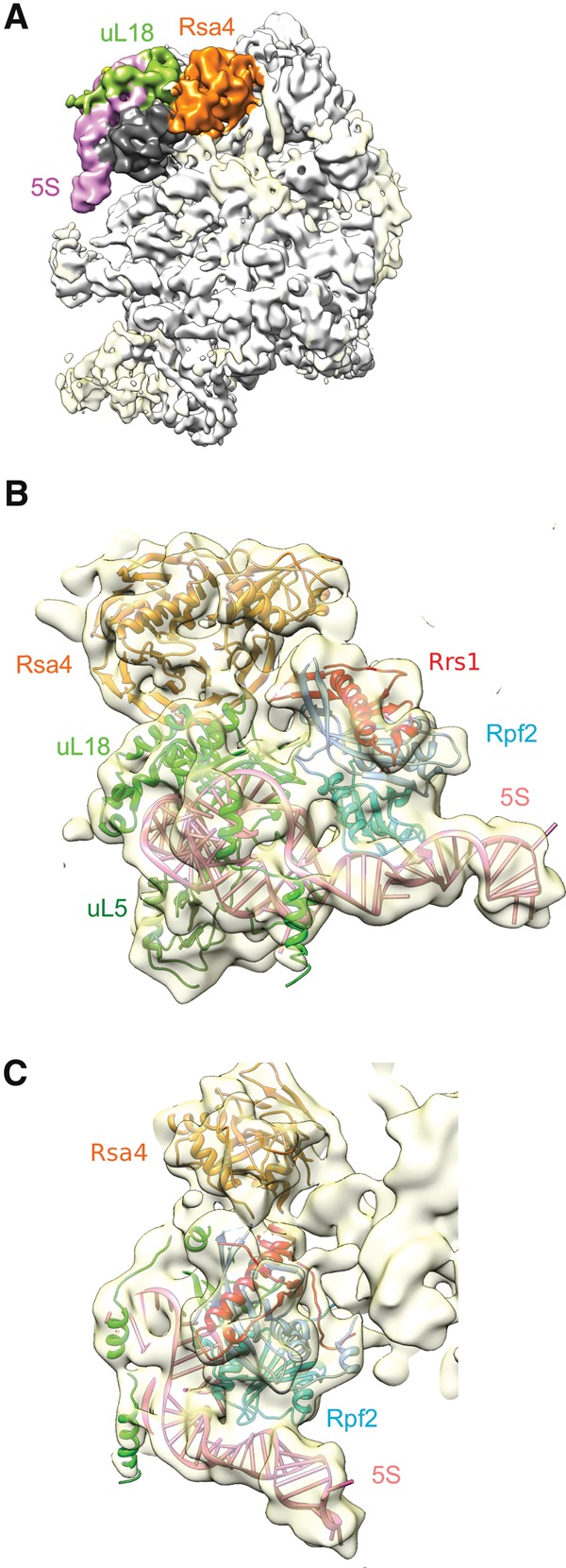
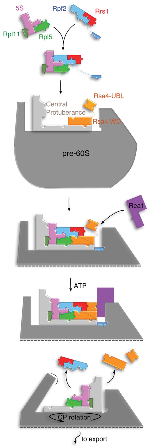
In eukaryotes, three of the four ribosomal RNAs (rRNAs)—the 5.8S, 18S, and 25S/28S rRNAs—are processed from a single pre-rRNA transcript and assembled into ribosomes. The fourth rRNA, the 5S rRNA, is transcribed by RNA polymerase III and is assembled into the 5S ribonucleoprotein particle (RNP), containing ribosomal proteins Rpl5/uL18 and Rpl11/uL5, prior to its incorporation into preribosomes. In mammals, the 5S RNP is also a central regulator of the homeostasis of the tumor suppressor p53. The nucleolar localization of the 5S RNP and its assembly into preribosomes are performed by a specialized complex composed of Rpf2 and Rrs1 in yeast or Bxdc1 and hRrs1 in humans. Here we report the structural and functional characterization of the Rpf2-Rrs1 complex alone, in complex with the 5S RNA, and within pre-60S ribosomes. We show that the Rpf2-Rrs1 complex contains a specialized 5S RNA E-loop-binding module, contacts the Rpl5 protein, and also contacts the ribosome assembly factor Rsa4 and the 25S RNA. We propose that the Rpf2-Rrs1 complex establishes a network of interactions that guide the incorporation of the 5S RNP in preribosomes in the initial conformation prior to its rotation to form the central protuberance found in the mature large ribosomal subunit.

Keywords: 5S RNP; Brix domain; p53; ribosome assembly.


Figures

Figure 1.
Figure 1.
Structure of the Rpf2–Rrs1 complex. (AC) Ribbon representation of three orthogonal views of the Rpf2–Rrs1 complex. The Brix internal duplicated (BID) domains are represented in light and dark blue, and Rrs1 is shown in red. (D) Superposition of the two Rpf2 BID domains shows that Rrs1 completes the structural elements in the second BID domain.
Figure 2.
Figure 2.
In vivo RNA-binding sites of Rpf2. (A,B) RNAs in Rpf2-HTP cells were UV cross-linked in cells growing in culture medium, trimmed and ligated to linkers, amplified by RT–PCR, and sequenced with an ionTorrent. Sequences were aligned with main hits to the 5S rRNA (nucleotides 1–120) (A) and the 25S rRNA (nucleotides 1–3396) (B). The frequency of recovery (hits per 100,000 mapped reads) is plotted for each individual nucleotide (shown in black). The locations of mutations/deletions that are likely due to RNA cross-linking to the residue are shown in red. The location of a classical contaminant sequence found in the 3′ end of the 25S rRNA (recovered with the “no tag” control experiment) is represented by a green bar. (C) Secondary structure of 5S RNA in yeast. The binding sites recovered for Rpf2-HTP are indicated on the sequence (blue). Mutated nucleotides that indicate a direct cross-link are indicated by red dots alongside the sequence. Known binding sites for 5S-binding protein TFIIIA, Rpl5, and Rpl11 are indicated as gray, light-green, and dark-green circles, respectively. (D) Secondary structure of the 25S RNA in yeast. The binding sites recovered for Rpf2-HTP are indicated on the sequence (blue). Mutated nucleotides are indicated by red dots alongside the sequence. Transient interaction between 5S and 25S in pre-60S particle is indicated as a gray square (Leidig et al. 2014).
Figure 3.
Figure 3.
In vitro interaction of the Rpf2–Rrs1 complex with 5S rRNA. (A) EMSA of the 5S rRNA. 5′-32P-labeled 5S rRNA was bound with 0, 4, 8, 16, 32, 64, 128, 256, 512, and 1024 nM indicated proteins. (B) Fractions of bound 5S rRNA for each protein concentration were quantified using PhosphoImager and plotted for the Rpf2/Rrs1 complex before (blue diamonds) or after (red triangles) 1 h of trypsin proteolysis. The theoretical curves are represented for the Rpf2/Rrs1 complex before (blue) and after (red) trypsin treatment. (CE) CD analysis of the RNA conformational changes upon Rpf2/Rrs1 binding. (C) CD spectra of the yeast 5S rRNA (yRNA) obtained when the protein complex Rpf2/Rrs1 and the yRNA are present in two separate cuvette compartments (dark green) or after mixing the two compartments (light green). (D) The same experiment as in C but with the protein complex Rpf2/Rrs1 after trypsin treatment. Spectrum were recorded before (dark purple) and after (light purple) mixing proteins and yRNA. (E) The same experiment as in C with bacterial 5S rRNA (bRNA) and the complex Rpf2/Rrs1 before (red) or after (orange) mixing the two compartments. (F) Filter-binding assay of the 5S rRNA E loop to the RPF2/RRS1 complex. The fluorescently labeled E loop of wild type or the G77U mutant was bound with 0, 25, 50, 100, 250, 1000, and 2000 nM protein complex. Fractions of the bound E loop of wild type or the G77U mutant of the 5S rRNA for each protein concentration were quantified using odyssey (Li-COR) and are plotted for the wild-type E loop (blue diamonds) and G77U mutant (red triangles). The theoretical curves are represented for the wild type(blue) and G77U mutant (red).
Figure 4.
Figure 4.
SAXS-derived solution structures of the Rpf2 subcomplexes. Solution structure model of the Rpf2–Rrs1 complex in the proteolyzed state (χ2 = 1.289) (A), the full-length Rpf2–Rrs1 complex (χ2 = 1.701) (B), the free 5S RNA (χ2 = 1.866) (C), and the Rpf2–Rrs1–5S complex with proteolyzed proteins (χ2 = 1.677). The structures are superposed to a representative envelope calculated by DAMMIN (A,B) or MONSA (C). The proteins and RNA are represented in the same orientation. The corresponding calculated X-ray scattering curves (dashed green) superposed to the experimental scattering curves (blue) are shown in the right panel. The locations of interesting 5S structural elements are indicated by their names to refer to Figure 2C.
Figure 5.
Figure 5.
Electron microscopy structure of pre-60S bound Rpf2–Rrs1. (A) Cryo-EM structure of the Alb1-TAP purified pre-60S particles (emd 2528) (Leidig et al. 2014). The unidentified density in contact with 5S (pink) and Rsa4 (orange) is shown in dark gray. (B,C) Orthogonal views of the Rpf2–Rrs1 crystal structure fit in the cryo-EM density. No optimization of the Rpf2–Rrs1 structure was performed.
Figure 6.
Figure 6.
RNA and protein contacts in the preribosomes. (A) Rpf2 binds the 5S RNA E loop. The 5S RNA and the Rpf2–Rrs1 complex were fitted simultaneously in the electron density with flexible molecular dynamics using MdFF. The E loop is colored in purple, and the looped-out guanine is depicted in a solid cartoon. (B) Rpf2 contacts to the 25S rRNA. The predicted C-terminal extension of Rpf2, not visible in the crystal structure, is shown by a dashed line. The rRNA sequence identified by CRAC is colored in orange. (C) Protein–protein interaction with the Rpf2–Rrs1 complex in the preribosome.
Figure 7.
Figure 7.
Jigsaw puzzle model for Rpf2/Rrs1 complex function in 5S RNP integration in preribosomes. Strong affinity of the Rpf2/Rrs1 complex for the 5S rRNA suggests that it recruits the free pool of 5S RNP. In this complex, the 5S RNP can only assemble the preribosomes containing the correct conformation. The preassembled Rpf2/Rrs1 targets the 5S RNP onto pre-60S ribosomes using interactions between Rpf2 and the 25S rRNA and between Rpf2/Rrs1/Rpl5 and Rsa4. The Rsa4 UBL domain is positioned by Rrs1 to allow the interaction with Rea1, which recycles Rsa4 and Rpf2/Rrs1 from the pre-60S particle and allows the 5S RNP to adopt its final configuration state through a 180° rotation of the CP.

References

    1. Adams PD, Afonine PV, Bunkóczi G, Chen VB, Davis IW, Echols N, Headd JJ, Hung L-W, Kapral GJ, Grosse-Kunstleve RW, et al. 2010. Phenix: a comprehensive Python-based system for macromolecular structure solution. Acta Crystallogr D Biol Crystallogr 66: 213–221. - PMC - PubMed
    1. Armistead J, Khatkar S, Meyer B, Mark BL, Patel N, Coghlan G, Lamont RE, Liu S, Wiechert J, Cattini PA, et al. 2009. Mutation of a gene essential for ribosome biogenesis, EMG1, causes Bowen-Conradi syndrome. Am J Hum Genet 84: 728–739. - PMC - PubMed
    1. Asano N, Kato K, Nakamura A, Komoda K, Tanaka I, Yao M. 2015. Structural and functional analysis of the Rpf2-Rrs1 complex in ribosome biogenesis. Nucleic Acids Res 43: 4746–4757. - PMC - PubMed
    1. Baßler J, Paternoga H, Holdermann I, Thoms M, Granneman S, Barrio-Garcia C, Nyarko A, Stier G, Clark SA, Schraivogel D, et al. 2014. A network of assembly factors is involved in remodeling rRNA elements during preribosome maturation. J Cell Biol 207: 481–498. - PMC - PubMed
    1. Bricogne G, Vonrhein C, Flensburg C, Schiltz M, Paciorek W. 2003. Generation, representation and flow of phase information in structure determination: recent developments in and around Sharp 2.0. Acta Crystallogr D Biol Crystallogr 59: 2023–2030. - PubMed

Publication types

MeSH terms

Associated data