. 2015 Aug 21;290(34):21054-21066.
doi: 10.1074/jbc.M115.642538. Epub 2015 Jul 9.

Dimerization Mediated by a Divergent Forkhead-associated Domain Is Essential for the DNA Damage and Spindle Functions of Fission Yeast Mdb1

Affiliations

Dimerization Mediated by a Divergent Forkhead-associated Domain Is Essential for the DNA Damage and Spindle Functions of Fission Yeast Mdb1

Shukun Luo et al. J Biol Chem. .

Abstract

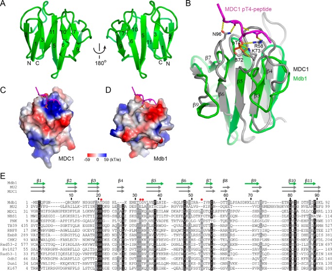
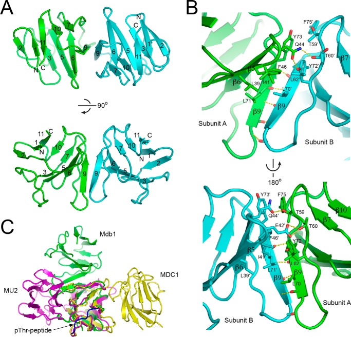
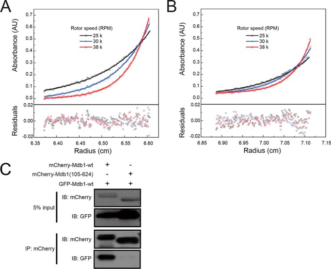
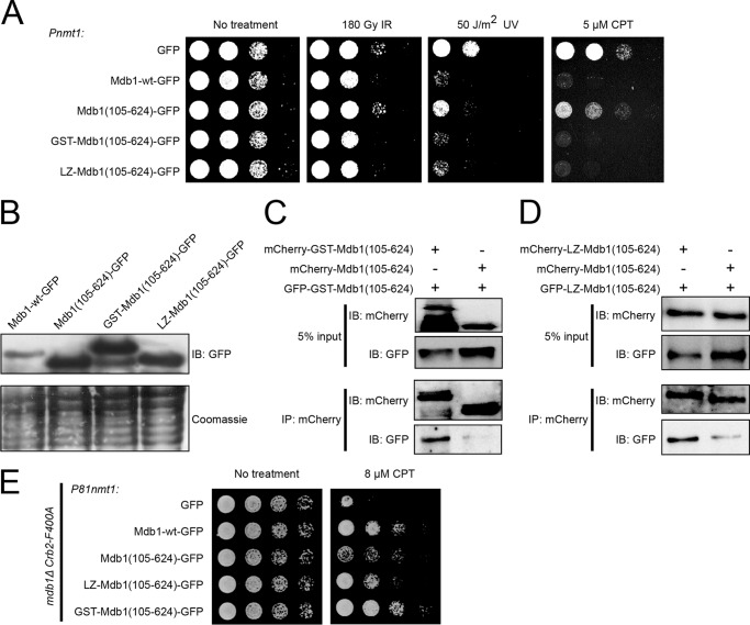
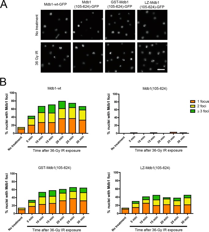
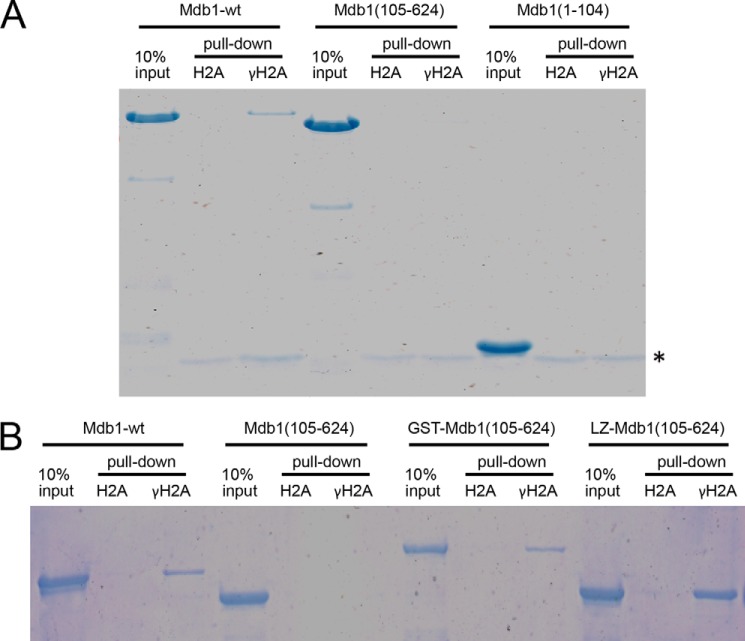
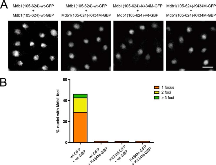
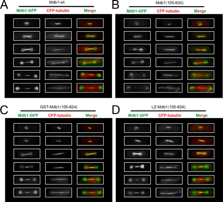
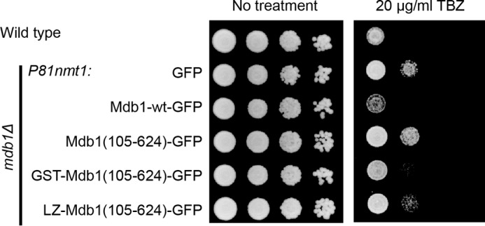
MDC1 is a key factor of DNA damage response in mammalian cells. It possesses two phospho-binding domains. In its C terminus, a tandem BRCA1 C-terminal domain binds phosphorylated histone H2AX, and in its N terminus, a forkhead-associated (FHA) domain mediates a phosphorylation-enhanced homodimerization. The FHA domain of the Drosophila homolog of MDC1, MU2, also forms a homodimer but utilizes a different dimer interface. The functional importance of the dimerization of MDC1 family proteins is uncertain. In the fission yeast Schizosaccharomyces pombe, a protein sharing homology with MDC1 in the tandem BRCA1 C-terminal domain, Mdb1, regulates DNA damage response and mitotic spindle functions. Here, we report the crystal structure of the N-terminal 91 amino acids of Mdb1. Despite a lack of obvious sequence conservation to the FHA domain of MDC1, this region of Mdb1 adopts an FHA-like fold and is therefore termed Mdb1-FHA. Unlike canonical FHA domains, Mdb1-FHA lacks all the conserved phospho-binding residues. It forms a stable homodimer through an interface distinct from those of MDC1 and MU2. Mdb1-FHA is important for the localization of Mdb1 to DNA damage sites and the spindle midzone, contributes to the roles of Mdb1 in cellular responses to genotoxins and an antimicrotubule drug, and promotes in vitro binding of Mdb1 to a phospho-H2A peptide. The defects caused by the loss of Mdb1-FHA can be rescued by fusion with either of two heterologous dimerization domains, suggesting that the main function of Mdb1-FHA is mediating dimerization. Our data support that FHA-mediated dimerization is conserved for MDC1 family proteins.

Keywords: DNA damage response; H2A histone family, member X (H2AFX); MDC1; Mdb1; Schizosaccharomyces pombe; crystal structure; dimerization; forkhead-associated domain (FHA domain); mitotic spindle; protein-protein interaction.


Figures

FIGURE 1.
FIGURE 1.
Domain organization of Mdb1. A, full-length Mdb1 protein was partially digested with trypsin and analyzed with SDS-PAGE and Coomassie staining. The migration positions of molecular makers are indicated. B, elution profile of Mdb1 digestion products in a Superdex 75 column. The elution positions of protein standards are indicated. C, peak fractions from gel filtration of trypsin-digested Mdb1 were analyzed with SDS-PAGE. D, domain diagram of Mdb1.
FIGURE 2.
FIGURE 2.
Structure of the FHA domain of Mdb1. A, ribbon representation of the FHA domain of Mdb1. Two opposite views are displayed. The β-strands are numbered according to structural equivalence to those of canonical FHA domains. The N and C termini are labeled. B, Mdb1-FHA lacks a Thr(P)-binding pocket. The structure of Mdb1-FHA is superimposed with the structure of MDC1-FHA bound with the MDC1-Thr(P)-4 peptide (Protein Data Bank code 3UNN). The interacting residues in the MDC1 FHA-phosphopeptide complex are shown in a stick and ball representation with phosphorus colored yellow, oxygen red, and nitrogen blue. Hydrogen bonds are shown as dashed lines. C and D, charge surfaces of the FHA domains of MDC1 (C) and Mdb1 (D). The surfaces are colored from blue to red for positively to negatively charged regions. The two structures have the same orientation as in B. The phosphopeptide ligand of MDC1 is displayed in the Mdb1 structure to indicate the region corresponding to the Thr(P)-binding pocket in MDC1. E, structure-based sequence alignment of FHA domains. Aligned are FHA domains from the following (Protein Data Bank codes are shown in parentheses): Mdb1 (4S3H), MU2 (3UV0), MDC1 (3UNN), NBS1 (3HUF), PNK (2W3O), TB39.8 (3POA), RNF8 (2PIE), EmbR (2FF4), CHK2 (1GXC), Rad53-FHA1 (1G6G), Rv1827 (2KFU), Rad53-FHA2 (1J4L), OdhI (2KB3), Dun1 (2JQL), and Ki67 (2AFF). Except for Mdb1 and MU2, all FHA domains bind a phosphopeptide in structure. The key phosphopeptide-binding residues are marked with red solid cycles. The secondary structures are indicated for FHA domains of Mdb1 (green arrows), MU2 (gray arrows), and MDC1 (gray arrows) above the sequences. Residue numbers are shown for Mdb1. Omitted residues are indicated by ∼. Residues that are conserved in at least 90 and 80% of these sequences are shaded in black and gray, respectively.
FIGURE 3.
FIGURE 3.
Dimeric structure of Mdb1-FHA. A, ribbon representation of Mdb1-FHA dimer structure shown in two orthogonal views. Subunits A and B are colored green and cyan, respectively. The β-strands are numbered as in Fig. 2, and the N and C termini are labeled. B, the dimer interface viewed from two opposite sides. Residues involved in dimerization are shown as sticks with oxygen colored red and nitrogen colored blue. Hydrogen bonds are shown as yellow dashed lines. C, one subunit of the FHA dimers of Mdb1 (green), MU2 (magenta), and MDC1 (yellow) are aligned. The bound Thr(P)-peptide of MDC1 is shown in blue.
FIGURE 4.
FIGURE 4.
Mdb1-FHA forms a stable dimer in vitro and is required for Mdb1 self-interaction in vivo. A and B, ultracentrifugation sedimentation equilibrium profiles of Mdb1 FHA domain. Proteins in concentrations of 67 μm (A) and 34 μm (B) were centrifuged at 25,000, 30,000, and 38,000 rpm. The curves are the best global fit of the distribution profiles against a self-association model with Kd = 375 nm. Fitting residuals are shown at the bottom. C, the FHA domain of Mdb1 is required for its self-interaction. mCherry-tagged Mdb1 was immunoprecipitated with RFP-trap agarose beads. The strains used were DY18067 and DY18069. IB, immunoblot.
FIGURE 5.
FIGURE 5.
FHA-mediated dimerization is important for the DNA damage functions of Mdb1. A, FHA-mediated dimerization is important for the genotoxin sensitivity induced by Mdb1 overexpression. Cells were pregrown in a thiamine-free medium for 20 h to allow full induction of the Pnmt1 promoter, and 5-fold serial dilutions were spotted on EMM-based thiamine-free plates. The strains used were DY15615, DY15616, DY17216, DY17218, and DY17220. B, the expression levels of different versions of Mdb1 analyzed in A. Coomassie staining of PVDF membrane after immunodetection was used to assess protein loading level and blotting efficiency (43). C, adding GST tag allowed Mdb1(105–624) to self-interact. The strains used were DY24566 and DY24567. D, adding LZ tag allowed Mdb1(105–624) to self-interact. The strains used were DY24570 and DY24571. E, FHA-mediated dimerization is required for the ability of Mdb1 to rescue the CPT sensitivity of mdb1Δ crb2-F400A double mutant. The expression was under the control of the P81nmt1 promoter. Cells were pregrown in a thiamine-free medium for 20 h before being spotted on CPT-containing thiamine-free plates. The strains used were DY16581, DY17124, DY17126, DY16579, and DY16573. IB, immunoblot.
FIGURE 6.
FIGURE 6.
FHA-mediated dimerization is important for the ability of Mdb1 to form nuclear foci. A, focus formation by Mdb1 requires FHA-mediated dimerization. Cells were imaged before and 30 min after exposure to 36 Gy of IR. The strains used were DY15603, DY15638, DY16309, and DY16307. Bar, 5 μm. B, a time course analysis of IR-induced Mdb1 focus formation. Approximately 200 nuclei were examined for each sample.
FIGURE 7.
FIGURE 7.
FHA-mediated dimerization is important for the ability of Mdb1 to interact with γH2A. A, the FHA domain is necessary but not sufficient for Mdb1 to bind γH2A. Recombinant Mdb1 proteins were expressed in bacteria and purified using the His6 tag. Biotinylated peptides that correspond to the C terminus of H2A.1, either unmodified (H2A) or phosphorylated on Ser-129 (γH2A), were incubated with the recombinant Mdb1 proteins. Peptides and associated proteins were pulled down by streptavidin Dynabeads and eluted by boiling in SDS-PAGE loading buffer. The eluates and 10% inputs were analyzed by SDS-PAGE followed by Coomassie staining. The asterisk indicates a background band released from the Dynabeads. B, adding a dimerization domain rescued the ability of Mdb1(105–624) to interact with γH2A. The experiment was performed as in A.
FIGURE 8.
FIGURE 8.
Mdb1 focus formation requires both subunits in a dimer to possess the γH2A-binding ability. A, co-expression of Mdb1(105–624)-GBP rescued the ability of Mdb1(105–624)-GFP to form nuclear foci, and this rescue was abolished when the K434M mutation was introduced into one or both proteins. Cells were imaged 30 min after exposure to 36 Gy of IR. The strains used were DY23263, DY24110, DY24112, and DY24114. Bar, 5 μm. B, quantitation of the results shown in A. Approximately 200 nuclei were examined for each sample.
FIGURE 9.
FIGURE 9.
The midzone accumulation of Mdb1 requires FHA-mediated dimerization. The strains used were DY15627, DY16639, DY16643, and DY16645. A, Mdb1-wt. B, Mdb1(105–624). C, GST-Mdb1(105–624). D, LZ-Mdb1(105–624).
FIGURE 10.
FIGURE 10.
FHA-mediated dimerization is important for the ability of Mdb1 to reverse the TBZ-resistant phenotype of mdb1Δ. Cells were pregrown in a thiamine-free medium for 20 h before being spotted on TBZ-containing thiamine-free plates. The strains used were LD327, DY15630, DY15603, DY15638, DY16309, and DY16307.

References

    1. Reinhardt H. C., Yaffe M. B. (2013) Phospho-Ser/Thr-binding domains: navigating the cell cycle and DNA damage response. Nat. Rev. Mol. Cell Biol. 14, 563–580 - PubMed
    1. Mermershtain I., Glover J. N. (2013) Structural mechanisms underlying signaling in the cellular response to DNA double strand breaks. Mutat. Res. 750, 15–22 - PMC - PubMed
    1. Rogakou E. P., Pilch D. R., Orr A. H., Ivanova V. S., Bonner W. M. (1998) DNA double-stranded breaks induce histone H2AX phosphorylation on serine 139. J. Biol. Chem. 273, 5858–5868 - PubMed
    1. Scully R., Xie A. (2013) Double strand break repair functions of histone H2AX. Mutat. Res. 750, 5–14 - PMC - PubMed
    1. Stucki M., Clapperton J. A., Mohammad D., Yaffe M. B., Smerdon S. J., Jackson S. P. (2005) MDC1 directly binds phosphorylated histone H2AX to regulate cellular responses to DNA double-strand breaks. Cell. 123, 1213–1226 - PubMed

Publication types

MeSH terms