Optical detection enhancement in porous volumetric microfluidic capture elements using refractive index matching fluids
- PMID: 26160546
- PMCID: PMC4516631
- DOI: 10.1039/c5an00988j
Optical detection enhancement in porous volumetric microfluidic capture elements using refractive index matching fluids
Abstract
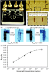
Porous volumetric capture elements in microfluidic sensors are advantageous compared to planar capture surfaces due to higher reaction site density and decreased diffusion lengths that can reduce detection limits and total assay time. However a mismatch in refractive indices between the capture matrix and fluid within the porous interstices results in scattering of incident, reflected, or emitted light, significantly reducing the signal for optical detection. Here we demonstrate that perfusion of an index-matching fluid within a porous matrix minimizes scattering, thus enhancing optical signal by enabling the entire capture element volume to be probed. Signal enhancement is demonstrated for both fluorescence and absorbance detection, using porous polymer monoliths in a silica capillary and packed beds of glass beads within thermoplastic microchannels, respectively. Fluorescence signal was improved by a factor of 3.5× when measuring emission from a fluorescent compound attached directly to the polymer monolith, and up to 2.6× for a rapid 10 min direct immunoassay. When combining index matching with a silver enhancement step, a detection limit of 0.1 ng mL(-1) human IgG and a 5 log dynamic range was achieved. The demonstrated technique provides a simple method for enhancing optical sensitivity for a wide range of assays, enabling the full benefits of porous detection elements in miniaturized analytical systems to be realized.
Figures


Similar articles
-
Flow-through microfluidic immunosensors with refractive index-matched silica monoliths as volumetric optical detection elements.Sens Actuators B Chem. 2018 Jan;254:878-886. doi: 10.1016/j.snb.2017.07.137. Epub 2017 Jul 21. Sens Actuators B Chem. 2018. PMID: 29225421 Free PMC article.
-
Impedimetric Immunosensing in a Porous Volumetric Microfluidic Detector.Sens Actuators B Chem. 2016 Oct 29;234:493-497. doi: 10.1016/j.snb.2016.05.015. Epub 2016 May 6. Sens Actuators B Chem. 2016. PMID: 27721569 Free PMC article.
-
A 3D porous polymer monolith-based platform integrated in poly(dimethylsiloxane) microchips for immunoassay.Analyst. 2013 May 7;138(9):2613-9. doi: 10.1039/c3an36744d. Analyst. 2013. PMID: 23478568
-
Fluid dynamics in capillary and chip electrochromatography.Electrophoresis. 2007 Feb;28(4):611-26. doi: 10.1002/elps.200600625. Electrophoresis. 2007. PMID: 17253632 Review.
-
Recent advances in optical fiber devices for microfluidics integration.J Biophotonics. 2016 Jan;9(1-2):13-25. doi: 10.1002/jbio.201500170. J Biophotonics. 2016. PMID: 27115035 Review.
Cited by
-
Biosensors Based on Inorganic Composite Fluorescent Hydrogels.Nanomaterials (Basel). 2023 May 26;13(11):1748. doi: 10.3390/nano13111748. Nanomaterials (Basel). 2023. PMID: 37299650 Free PMC article. Review.
-
Flow-through microfluidic immunosensors with refractive index-matched silica monoliths as volumetric optical detection elements.Sens Actuators B Chem. 2018 Jan;254:878-886. doi: 10.1016/j.snb.2017.07.137. Epub 2017 Jul 21. Sens Actuators B Chem. 2018. PMID: 29225421 Free PMC article.
-
Impedimetric Immunosensing in a Porous Volumetric Microfluidic Detector.Sens Actuators B Chem. 2016 Oct 29;234:493-497. doi: 10.1016/j.snb.2016.05.015. Epub 2016 May 6. Sens Actuators B Chem. 2016. PMID: 27721569 Free PMC article.
-
Novel functionalities of hybrid paper-polymer centrifugal devices for assay performance enhancement.Biomicrofluidics. 2017 Sep 12;11(5):054101. doi: 10.1063/1.5002644. eCollection 2017 Sep. Biomicrofluidics. 2017. PMID: 28966698 Free PMC article.
References
-
- American Academy of Microbiology. Bringing the Lab to the Patient: Developing Point-of-Care Diagnostics for Resource Limited Settings. 2011
-
- Chin CD, Linder V, Sia SK. Lab Chip. 2012;12:2118–2134. - PubMed
-
- Yager P, Domingo GJ, Gerdes J. Annu. Rev. Biomed. Eng. 2008;10:107–144. - PubMed
-
- Ellerbee AK, Phillips ST, Siegel AC, Mirica Ka, Martinez AW, Striehl P, Jain N, Prentiss M, Whitesides GM. Anal. Chem. 2009;81:8447–8452. - PubMed
Publication types
MeSH terms
Substances
Grants and funding
LinkOut - more resources
Full Text Sources
Other Literature Sources

