. 2016 Jan 20;24(3):141-56.
doi: 10.1089/ars.2015.6272. Epub 2015 Aug 17.

Redox Activation of the Universally Conserved ATPase YchF by Thioredoxin 1

Affiliations

Redox Activation of the Universally Conserved ATPase YchF by Thioredoxin 1

Liya Hannemann et al. Antioxid Redox Signal. .

Abstract

Aims: YchF/Ola1 are unconventional members of the universally conserved GTPase family because they preferentially hydrolyze ATP rather than GTP. These ATPases have been associated with various cellular processes and pathologies, including DNA repair, tumorigenesis, and apoptosis. In particular, a possible role in regulating the oxidative stress response has been suggested for both bacterial and human YchF/Ola1. In this study, we analyzed how YchF responds to oxidative stress and how it potentially regulates the antioxidant response.

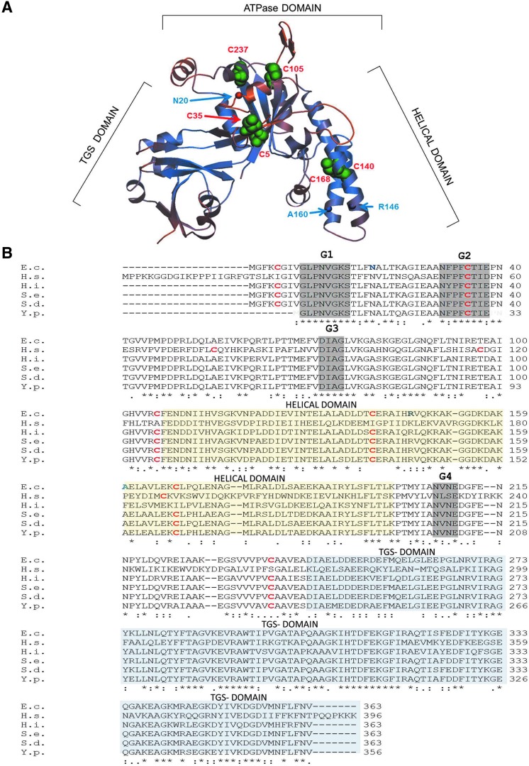
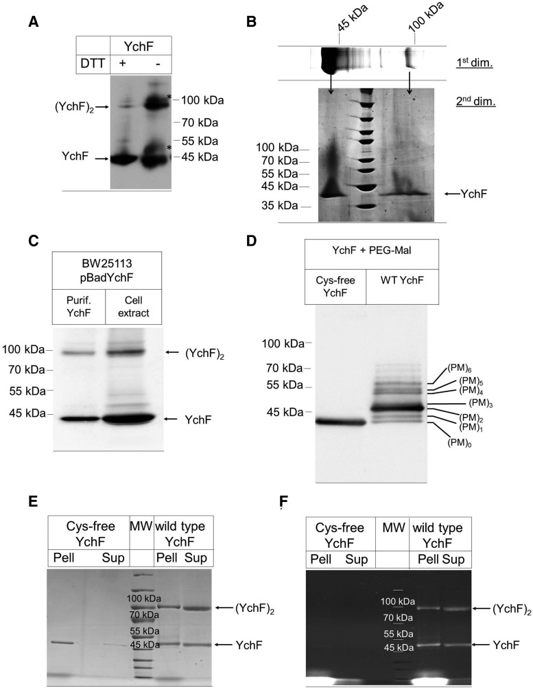
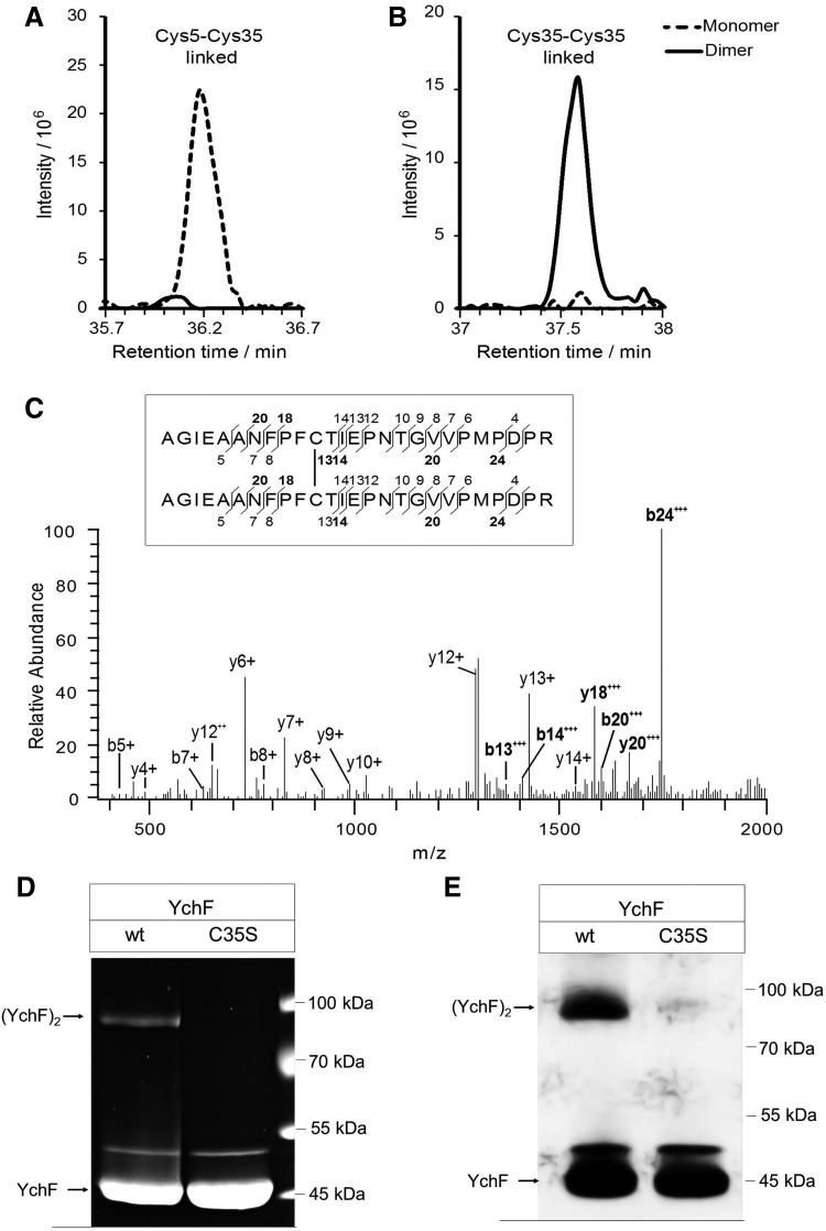
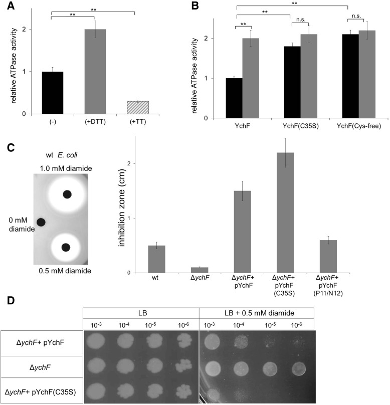
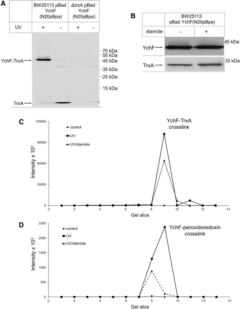
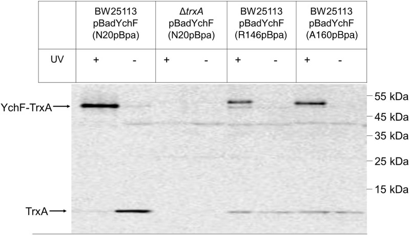
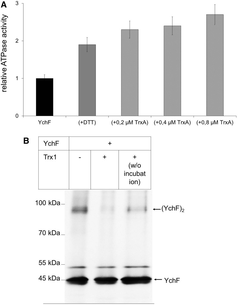
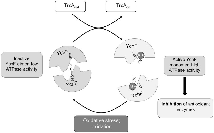
Results: Our data identify a redox-regulated monomer-dimer equilibrium of YchF as a key event in the functional cycle of YchF. Upon oxidative stress, the oxidation of a conserved and surface-exposed cysteine residue promotes YchF dimerization, which is accompanied by inhibition of the ATPase activity. No dimers were observed in a YchF mutant lacking this cysteine. In vitro, the YchF dimer is dissociated by thioredoxin 1 (TrxA) and this stimulates the ATPase activity. The physiological significance of the YchF-thioredoxin 1 interaction was demonstrated by in vivo cross-linking, which validated this interaction in living cells. This approach also revealed that both the ATPase domain and the helical domain of YchF are in contact with TrxA.

Innovation: YchF/Ola1 are the first redox-regulated members of the universally conserved GTPase family and are inactivated by oxidation of a conserved cysteine residue within the nucleotide-binding motif.

Conclusion: Our data provide novel insights into the regulation of the so far ill-defined YchF/Ola1 family of proteins and stipulate their role as negative regulators of the oxidative stress response.


Figures

<b>FIG. 1.</b>
FIG. 1.
Position and sequence conservation of cysteine residues in YchF/Ola1. (A) The crystal structure of the YchF homologue of Haemophilus influenzae (1JAL) was used to model the probable position of the six cysteine residues in Escherichia coli YchF using the Swiss Model (http://swissmodel.expasy.org/interactive/kFyFA3/models). Cysteine residues are labeled with green spheres and the amino acid position based on the E. coli numbering is indicated in red. In addition, the residues where the UV-induced cross-linker pBpa was incorporated are indicated by circles and the amino acid position based on the E. coli numbering is indicated in blue. (B) A sequence alignment of YchF homologues from different enterobacteria was generated by using the Clustal 2.0 software and compared with the H. influenzae YchF and Homo sapiens Ola1 sequence. Cysteine residues are shown in red and bold and the positions where pBpa was incorporated in the E. coli YchF are shown in blue and bold. The G1-G4 motifs of the nucleotide-binding site are boxed in gray, the helical domain is boxed in yellow, and the TGS domain in blue. E.c., Escherichia coli; H.s., Homo sapiens; H.i., Haemophilus influenzae; S.e., Salmonella enterica; Y.p., Yersinia pestis; S.d., Shigella dysentericae. To see this illustration in color, the reader is referred to the web version of this article at www.liebertpub.com/ars
<b>FIG. 2.</b>
FIG. 2.
YchF forms redox- and cysteine-dependent physiological dimers. (A) Purified YchF was denatured at 37°C for 15 min at either reducing (+25 mM DTT) or nonreducing conditions (−DTT) and after sodium dodecyl sulfate–polyacrylamide gel electrophoresis (SDS-PAGE) and Western transfer probed with α-YchF antibodies. Two weaker bands were also recognized by α-YchF antibodies (*) in the absence of DTT. (B) Purified YchF was separated on SDS-PAGE under nonreducing conditions as in A (1st dim.), and after Coomassie staining, the YchF monomer and dimer bands were excised out of the gel and separated on a second dimension SDS-PAGE (2nd dim.) under reducing conditions. (C) Purified YchF and lysozyme-lysed E. coli cells expressing YchF were separated under nonreducing conditions on SDS-PAGE and YchF was detected after Western transfer with α-YchF antibodies. (D) Purified wild-type YchF (wt YchF) and a cysteine-free derivative (Cys-free YchF) were reduced and incubated with the thiol-modifying agent, PEG-Mal, and subsequently separated by SDS-PAGE. After Western transfer, the membrane was probed with α-YchF antibodies. (PM)0–6 reflect the number of labeled cysteines. (E, F) Purified Cys-free and wild-type YchF were incubated with the fluorescent thiol-modifying agent fluorescein-5-maleimide. Subsequently, pellet (Pell) and supernatant (Sup) fractions were prepared and samples separated by SDS-PAGE under nonreducing conditions, followed by Coomassie staining (E) or in-gel fluorescence analysis (F).
<b>FIG. 3.</b>
FIG. 3.
Identification and quantification of disulfide-linked peptides in YchF monomer and dimer. (A) Extracted ion chromatograms of the Cys5-Cys35-linked peptides, CGIVGLPNVGK and AGIEAANFPFCTIEPNTGVVPM*PDPR (precursor m/z 953.9757, charge +4), determined by liquid chromatography–mass spectrometry analyses of the monomer (dashed line) and dimer band (solid line). (B) Equivalent to A, but for the Cys35-Cys35-linked peptide AGIEAANFPFCTIEPNTGVVPM*PDPR (precursor m/z 1379.6581, charge +4). (C) Fragment spectrum of the Cys35-Cys35 disulfide-linked peptide AGIEAANFPFCTIEPNTGVVPM*PDPR. b- and y-type fragment ions are annotated in the mass spectrum and in the sequence (inset). Fragments containing the disulfide bridge are shown in bold. (D) Wild-type YchF and YchF containing a single cysteine-to-serine replacement at position 35 (YchFC35S) were purified and separated by SDS-PAGE under nonreducing conditions. Proteins were subsequently stained with Coomassie. (E) As in D, but samples were probed with α-YchF antibodies after Western blotting. M*, oxidized methionine.
<b>FIG. 4.</b>
FIG. 4.
Dimerization of YchF inhibits its ATPase activity (A) The ATPase activity of purified wild-type YchF (black bar) was determined by measuring phosphate release of γ-33P-labeled ATP. When indicated, YchF was preincubated with either 10 mM DTT (gray bar) or 10 mM tetrathionate (TT) (hatched bar) for 5 min at room temperature before measuring the activity. A relative YchF activity of 1 corresponds to ∼0.35 nmol ATP/(min × mg protein). The relative ATPase activities shown are the mean ± SD (n > 3). **p < 0.01. (B) The ATPase activities of wild-type YchF, the YchF(C35S) mutant, and of the cysteine-free YchF mutant were determined without (black bars) or after preincubation with 10 mM DTT (gray bars). A relative YchF activity of 1 corresponds to ∼0.35 nmol ATP/(min × mg protein). The relative ATPase activities shown are the mean ± SD (n = 3). **p < 0.01; n.s. not significant. (C) E. coli strains were adjusted to an optical density of 0.5, mixed with Top agar, and poured on LB Plates. Filter discs were soaked with different diamide concentrations and placed on these plates. Inhibition zones were quantified after ∼5 h of growth and the results for wild-type E. coli are shown (left panel). Quantification of two independent experiments in the presence of 0.5 mM diamide (right panel). pYchF(P11/N12) corresponds to an ATPase-deficient YchF derivative. (D) The indicated E. coli strains were grown overnight on liquid LB medium without antibiotics and adjusted to an optical density of 1.0 before serial dilution. Dilutions were then spotted onto LB plates or LB plates containing 0.5 mM diamide. Plates were incubated at 37°C and analyzed after ∼12 h of growth.
<b>FIG. 5.</b>
FIG. 5.
YchF cross-links to thioredoxin in vivo. (A) Wild-type E. coli cells or a ΔtrxA E. coli strain expressing YchF with the UV-dependent cross-linker pBpA incorporated at position N20 were either UV exposed (+) or kept in the dark (−). After cross-linking, YchF was purified, separated by SDS-PAGE, and after Western transfer probed with α-TrxA antibodies. (B) Wild-type E. coli cells expressing YchF(N20pBpa) were treated with 50 μM diamide and TCA precipitated. The pellet after centrifugation was separated by SDS-PAGE and after Western transfer probed with either α-YchF or with α-TrxA antibodies, (C, D) YchF(N20pBpa) cross-linking products were separated by SDS-PAGE, individual gel slices corresponding to the MW range of 130 to 40 kDa were excised, subjected to in-gel digestion using trypsin, and analyzed by mass spectrometry. Data were analyzed for peptides derived from TrxA (C) and peroxiredoxin (D). Shown are summed peptide intensities measured in samples obtained without UV exposure (noncross-linked, ♦, dotted line), after UV exposure (cross-linked, ■, solid line), and after UV exposure of diamide-treated cells (▲, dashed line).
<b>FIG. 6.</b>
FIG. 6.
Thioredoxin interacts with both the N-terminus and the helical domain of YchF in vivo. In vivo cross-linking was performed as in Fig. 5 with pBpa inserted at position N20, R146, or N160. Samples were subsequently analyzed by Western blotting using α-TrxA antibodies. Cross-linked products of positions, R146 and 160, migrated in two distinct species, which is a common phenomenon of pBpa cross-linking (25, 31, 38) and probably reflects different three-dimensional structures.
<b>FIG. 7.</b>
FIG. 7.
Thioredoxin increases the ATPase activity of YchF and reduces the YchF dimer. (A) Purified YchF (0.2 μM) was incubated with either DTT (10 mM) (gray bar) or increasing TrxA concentrations (hatched bars) and the ATPase activity was measured as in Fig. 4. The activity of wild-type YchF was set to one and corresponds to 0.35 nmol ATP/(min × mg protein). The values observed were corrected for the background ATP hydrolysis detected in the purified TrxA sample. (B) Purified YchF was incubated for 30 min at 37°C in the presence or absence of purified TrxA in a 1:1 molar ratio. As a control, TrxA was added to YchF without further incubation (w/o incubation). Samples were analyzed by SDS-PAGE under nonreducing conditions and probed with α-YchF antibodies after Western transfer.
<b>FIG. 8.</b>
FIG. 8.
Model for the redox regulation of YchF, a universally conserved inhibitor of the oxidative stress response. YchF acts as inhibitor of the oxidative stress response in both E. coli (53) and H. sapiens (56, 57). Oxidative stress regulates YchF expression and activity at both the transcriptional level (53) and the post-translational level. In the absence of oxidative stress, YchF exists primarily as a monomer with high ATPase activity, which is required for the inhibition of antioxidant enzymes. Upon oxidative stress, cysteine residue 35, which is located within the G2 motif of the ATPase domain, is oxidized, leading to YchF dimer formation, which prevents ATP hydrolysis. YchF dimerization upon oxidative stress probably includes an intramolecular disulfide bridge between cysteine 5 and cysteine 35 as an intermediate. Dimerization and concomitant ATPase inhibition alleviate the inhibition of antioxidant enzymes. If oxidative stress eases, YchF dimers are dissociated by the activity of thioredoxin 1 (TrxA).

References

    1. Akhova AV. and Tkachenko AG. ATP/ADP alteration as a sign of the oxidative stress development in Escherichia coli under antibiotic treatment. FEMS Microbiol Lett 353: 69–76, 2014 - PubMed
    1. Alexander RW. and Schimmel P. Domain-domain communication in aminoacyl-tRNA synthetases. Prog Nucleic Acid Res Mol Biol 69: 317–349, 2001 - PubMed
    1. Antelmann H. and Helmann JD. Thiol-based redox switches and gene regulation. Antioxid Redox Signal 14: 1049–1063, 2011 - PMC - PubMed
    1. Aslund F, Zheng M, Beckwith J, and Storz G. Regulation of the OxyR transcription factor by hydrogen peroxide and the cellular thiol-disulfide status. Proc Natl Acad Sci U S A 96: 6161–6165, 1999 - PMC - PubMed
    1. Baker LM. and Poole LB. Catalytic mechanism of thiol peroxidase from Escherichia coli. Sulfenic acid formation and overoxidation of essential CYS61. J Biol Chem 278: 9203–9211, 2003 - PMC - PubMed

Publication types

MeSH terms

LinkOut - more resources