

Isolated pancreatic cysticercal cyst presenting as a diagnostic challenge: diagnosis and treatment review
- PMID: 26160552
- PMCID: PMC4499691
- DOI: 10.1136/bcr-2015-210774
Isolated pancreatic cysticercal cyst presenting as a diagnostic challenge: diagnosis and treatment review
Abstract
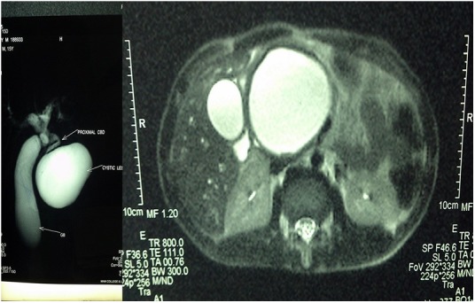
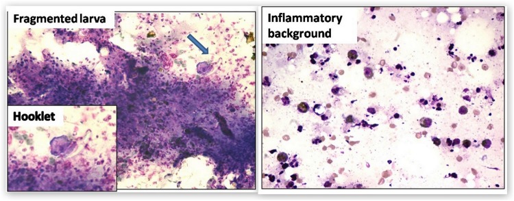
Human cysticercosis most commonly affects the subcutaneous tissues, skeletal muscles, lungs, brain, eyes, liver and, rarely, the heart, thyroid and pancreas. Owing to vague clinical presentation and unfamiliarity of clinicians with this entity, it is difficult to diagnosis when seen as an isolated cyst. We present a case of a 16-year-old boy who presented with an upper abdominal lump and jaundice. Ultrasonography (USG) and MRI of the abdomen were carried out, which revealed a cystic mass (8.5 × 7 × 7 cm) in the pancreas. No evidence of solid component or papillary projections was noted within the lesion. Tumour markers carcinoembryonic antigen (CEA) and cancer antigen (CA 19-9) were normal. Fine needle aspiration cytology was performed, which revealed the presence of cysticercus larvae, along with a foreign body giant cell reaction. The patient was treated with therapeutic aspiration and antihelminthic therapy. Since then, he has been symptom free and under regular follow-up for the last 1 year. A diagnosis of cysticercal cyst at atypical sites is very rare and depends mainly on histopathological examination, which, along with USG and MRI, can give an accurate analysis. These cysts can be very well treated non-surgically with antihelminthics and aspiration.
2015 BMJ Publishing Group Ltd.
Figures
Similar articles
-
Lymphoepithelial cysts of the pancreas. Can preoperative imaging distinguish this benign lesion from malignant or pre-malignant cystic pancreatic lesions?JOP. 2013 May 10;14(3):250-5. doi: 10.6092/1590-8577/1229. JOP. 2013. PMID: 23669473
-
Cyst wall puncture and aspiration during EUS-guided fine needle aspiration may increase the diagnostic yield of mucinous cysts of the pancreas.J Clin Gastroenterol. 2011 Feb;45(2):164-9. doi: 10.1097/MCG.0b013e3181eed6d2. J Clin Gastroenterol. 2011. PMID: 20818233
-
Cystic lesions of the pancreas. A diagnostic and management dilemma.Pancreatology. 2008;8(3):236-51. doi: 10.1159/000134279. Epub 2008 May 23. Pancreatology. 2008. PMID: 18497542 Review.
-
Molecular analysis of cyst fluid aspiration in the diagnosis and risk assessment of cystic lesions of the pancreas.Clin Transl Sci. 2012 Feb;5(1):102-7. doi: 10.1111/j.1752-8062.2011.00312.x. Epub 2011 Dec 8. Clin Transl Sci. 2012. PMID: 22376266 Free PMC article. Review.
References
-
- Evans CAW, Garcia HH, Gilman RH. Cysticercosis. In: Strickland GT, ed Hunter's tropical medicine. 8th edn Philadelphia, PA: WB Saunders Co, 2000:862.
-
- Krishnaswami CS. Case of cysticercuscellulose. Ind Med Gaz 1912;27:43–4. - PubMed
Publication types
MeSH terms
Substances
LinkOut - more resources
Full Text Sources
Other Literature Sources
Medical