

Delayed mirror visual feedback presented using a novel mirror therapy system enhances cortical activation in healthy adults
- PMID: 26160599
- PMCID: PMC4498534
- DOI: 10.1186/s12984-015-0053-1
Delayed mirror visual feedback presented using a novel mirror therapy system enhances cortical activation in healthy adults
Abstract
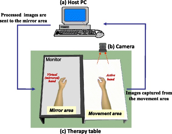
Background: Mirror visual feedback (MVF) generated in mirror therapy (MT) with a physical mirror promotes the recovery of hemiparetic limbs in patients with stroke, but is limited in that it cannot provide an asymmetric mode for bimanual coordination training. Here, we developed a novel MT system that can manipulate the MVF to resolve this issue. The aims of this pilot study were to examine the feasibility of delayed MVF on MT and to establish its effects on cortical activation in order to understand how it can be used for clinical applications in the future.
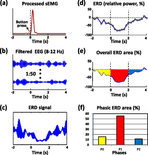
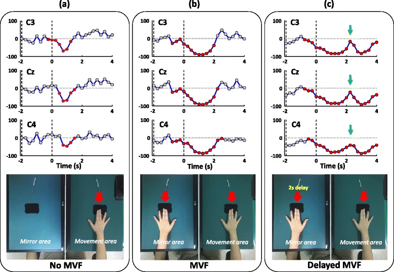
Methods: Three conditions (no MVF, MVF, and 2-s delayed MVF) presented via our digital MT system were evaluated for their time-course effects on cortical activity by event-related desynchronization (ERD) of mu rhythm electroencephalography (EEG) during button presses in 18 healthy adults. Phasic ERD areas, defined as the areas of the relative ERD curve that were below the reference level and within -2-0 s (P0), 0-2 s (P1), and 2-4 s (P2) of the button press, were used.
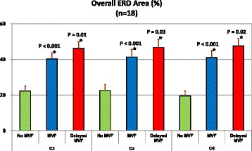
Results: The overall (P0 to P2) and phasic ERD areas were higher when MVF was provided compared to when MVF was not provided for all EEG channels (C3, Cz, and C4). Phasic ERD areas in the P2 phase only increased during the delayed-MVF condition. Significant enhancement of cortical activation in the mirror neuron system and an increase in attention to the unseen limb may play major roles in the response to MVF during MT. In comparison to the no MVF condition, the higher phasic ERD areas that were observed during the P1 phase in the delayed-MVF condition indicate that the image of the still hand may have enhanced the cortical activation that occurred in response to the button press.
Conclusions: This study is the first to achieve delayed MVF for upper-limb MT. Our approach confirms previous findings regarding the effects of MVF on cortical activation and contributes additional evidence supporting the use of this method in the future for upper-limb motor training in patients with stroke.
Figures
Similar articles
-
Enhancing mirror visual feedback with intermittent theta burst stimulation in healthy adults.Restor Neurol Neurosci. 2019;37(5):483-495. doi: 10.3233/RNN-190927. Restor Neurol Neurosci. 2019. PMID: 31424421
-
Neurophysiological effects of mirror visual feedback in stroke patients with unilateral hemispheric damage.Brain Res. 2018 Dec 1;1700:170-180. doi: 10.1016/j.brainres.2018.09.003. Epub 2018 Sep 5. Brain Res. 2018. PMID: 30194016
-
Alternative Motor Task-Based Pattern Training With a Digital Mirror Therapy System Enhances Sensorimotor Signal Rhythms Post-stroke.Front Neurol. 2019 Nov 22;10:1227. doi: 10.3389/fneur.2019.01227. eCollection 2019. Front Neurol. 2019. PMID: 31824406 Free PMC article.
-
The mirror neuron system and treatment of stroke.Dev Psychobiol. 2012 Apr;54(3):293-310. doi: 10.1002/dev.20504. Epub 2010 Nov 24. Dev Psychobiol. 2012. PMID: 22415917 Review.
-
ERD/ERS patterns reflecting sensorimotor activation and deactivation.Prog Brain Res. 2006;159:211-22. doi: 10.1016/S0079-6123(06)59014-4. Prog Brain Res. 2006. PMID: 17071233 Review.
Cited by
-
Mirror and Vibration Therapies Effects on the Upper Limbs of Hemiparetic Patients after Stroke: A Pilot Study.Rehabil Res Pract. 2018 Nov 4;2018:6183654. doi: 10.1155/2018/6183654. eCollection 2018. Rehabil Res Pract. 2018. PMID: 30519490 Free PMC article.
-
Associated Mirror Therapy Enhances Motor Recovery of the Upper Extremity and Daily Function after Stroke: A Randomized Control Study.Neural Plast. 2021 Sep 29;2021:7266263. doi: 10.1155/2021/7266263. eCollection 2021. Neural Plast. 2021. PMID: 34630560 Free PMC article. Clinical Trial.
-
EEG Based Analysis of Cortical Activity during Mirror Visual Feedback Target-Directed Movement.Annu Int Conf IEEE Eng Med Biol Soc. 2019 Jul;2019:5156-5159. doi: 10.1109/EMBC.2019.8857945. Annu Int Conf IEEE Eng Med Biol Soc. 2019. PMID: 31947019 Free PMC article.
-
Neural Processes Underlying Mirror-Induced Visual Illusion: An Activation Likelihood Estimation Meta-Analysis.Front Hum Neurosci. 2020 Jul 31;14:276. doi: 10.3389/fnhum.2020.00276. eCollection 2020. Front Hum Neurosci. 2020. PMID: 32848663 Free PMC article.
-
The Effects of Mirror Feedback during Target Directed Movements on Ipsilateral Corticospinal Excitability.Front Hum Neurosci. 2017 May 11;11:242. doi: 10.3389/fnhum.2017.00242. eCollection 2017. Front Hum Neurosci. 2017. PMID: 28553218 Free PMC article.
References
Publication types
MeSH terms
LinkOut - more resources
Full Text Sources
Other Literature Sources
Medical
Research Materials
Miscellaneous
