Review
. 2015 Aug;16(8):923-38.
doi: 10.15252/embr.201540434. Epub 2015 Jul 9.

Presynaptic active zones in invertebrates and vertebrates

Affiliations
Review

Presynaptic active zones in invertebrates and vertebrates

Frauke Ackermann et al. EMBO Rep. 2015 Aug.

Abstract

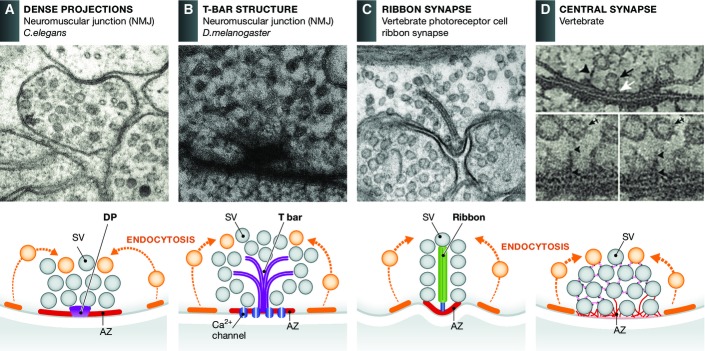
The regulated release of neurotransmitter occurs via the fusion of synaptic vesicles (SVs) at specialized regions of the presynaptic membrane called active zones (AZs). These regions are defined by a cytoskeletal matrix assembled at AZs (CAZ), which functions to direct SVs toward docking and fusion sites and supports their maturation into the readily releasable pool. In addition, CAZ proteins localize voltage-gated Ca(2+) channels at SV release sites, bringing the fusion machinery in close proximity to the calcium source. Proteins of the CAZ therefore ensure that vesicle fusion is temporally and spatially organized, allowing for the precise and reliable release of neurotransmitter. Importantly, AZs are highly dynamic structures, supporting presynaptic remodeling, changes in neurotransmitter release efficacy, and thus presynaptic forms of plasticity. In this review, we discuss recent advances in the study of active zones, highlighting how the CAZ molecularly defines sites of neurotransmitter release, endocytic zones, and the integrity of synapses.

Keywords: active zone; cytoskeletal matrix; fusion; release; synaptic vesicle.


Figures

Figure 1
Figure 1
Active zones display different morphologies Electron micrographs and schematic drawings reveal that AZs can be divided into morphologically distinct groups: those with elaborate electron-dense projections such as T-bars and ribbons and those with less prominent dense projections including C. elegans active zones and those present at most vertebrate central nervous system synapses. (A) AZ from a C. elegans NMJ. In general, these AZs are quite simple and generally characterized as a broad surface of plasma membrane situated between electron-dense projections (DPs) and flanking cellular tight junctions. (B) AZs present at the NMJ of the fly Drosophila melanogaster. These are more elaborate forming a platform consisting of a meshwork of filaments overlaying a pedestal which gave them the name T-bars. (C) Vertebrate photoreceptor cell ribbon synapse. These synapses are characterized by a large AZ with a specialized organelle, the synaptic ribbon, which tethers large numbers of SVs near the AZ, facilitating fast-sustained synaptic transmission. (D) AZs of vertebrate central synapses are less complex than at sensory synapses, exhibiting fine filamentous projections that connect proximal (docked vesicles) and more distally located SVs, up to 100 nm, to the plasma membrane holding them close to the release sites. In schematic drawing, active and endocytic (not shown in EM micrograph) zones are marked as red and orange, respectively. Reproduced with permission, from (A); Hollmann and Sigrist (B); (C); (D).
Figure 2
Figure 2
Cytoskeletal matrix proteins organize synaptic vesicle release sites at presynaptic active zones (A) 3D reconstruction of filaments (pink) and SVs (yellow) tethered near the active zone from a vertebrate (rat) hippocampal synapse. Docked vesicles are in blue. Reproduced with permission from . (B) Schematic diagram of CAZ molecules mediating the capture (synapsin, actin), docking (SNARE complex), priming (RIM, Munc13, Rab3) and fusion (synaptotagmin) of SVs and VGCCs (Bassoon, RBP, RIM, ELKS) at presynaptic active zones. At present, the spatial relationship of these molecules within the cryo-fixed EM image in (A) is not well resolved.
Figure 3
Figure 3
Macromolecular organization of the frog NMJ At the NMJ of the frog, SVs are arranged in highly organized linear arrays. The precise alignment of the vesicles is achieved through different filamentous structures, which contact the vesicles. (A) Electron micrograph showing subdomain organization of SVs at the frog NMJ. Synaptic vesicles are held in place through filamentous structures. Scale bar, 50 nm. (B) Schematic representation of the electron micrograph in (A) with summed outlined of AZ material (AZM) macromolecules colored. (C) 10-nm-thick surface model of AZM and SVs shown in (A) and (B), derived from eight adjacent slices. Docked vesicles close to the plasma membrane are attached to several filaments contrary to vesicles further away that are only connected to one filament. (D) 25-nm-thick surface model of an AZ, capturing a former docked vesicle that has fused with the plasma membrane but remains in contact with ribs, spars, and boom implying that AZM participates in SV fusion. (E) Surface model, ∼10 nm thick, showing in 3D the nubs linked by transmembrane bands connected to rib, booms, and spars. (F) Schematic diagram of the luminal assembly of macromolecules within a docked SV that form contact with rib, boom, spar, and pin as well as non-AZM molecules. (G) Composition diagram of layers of AZM shown in transverse plane of the AZ at frog NMJ. The main body of AZM includes beams, ribs, and pegs; the intermediate layer: steps and spars; and the deep layer masts, booms, and topmasts. (H) Diagram of NMJ revealing the repetitive array of core AZ proteins centered around a mast (green) that contacts surrounding SV through ribs, spars booms and topmasts. This geometric arrangement allows the creation of tens of SV docking and fusion sites. Reproduced with permission from Szule and Harlow ,.
Figure 4
Figure 4
Model of presynaptic bouton and active zone organization A section through the active and endocytic zones of a vertebrate synapse indicating the spatial distribution and copy number of presynaptic proteins that help define the presynaptic AZ as the site of SV exocytosis and clathrin-mediated endocytosis. Panel at the bottom, a graphical legend of the predicted structures of presynaptic proteins included in the model. Displayed SVs have a diameter of 42 nm. The image was generously created by Burkhard Rammner in the Rizzoli Laboratory. See also .
Figure 5
Figure 5
Synaptic vesicle cycle at the presynaptic terminal The presynaptic AZ functionally defines the space within boutons where upon calcium influx synaptic vesicle fusion and neurotransmission takes place (lower panel). It is the center of the SV life cycle. Vesicles are recruited from the vesicle cluster toward the AZ where they undergo maturation steps such as docking and priming and finally fuse with the plasma membrane upon action potential stimulation. After exocytosis, SV protein and membrane retrieval occurs through endocytosis in a region spatially adjacent to the active zone, the peri-active zone. It is an important compensatory reaction to recapture excess membrane and generate new SVs. Different endocytosis pathways are known and can be visualized by EM following evoke stimulation (top panels). Clathrin-mediated endocytosis (A) is a slow form of membrane retrieval. Synaptic vesicle proteins and membrane are taken up through the formation of clathrin-coated pits, which are later pinched off from the membrane through the GTPase dynamin. After uncoating, the newly formed vesicles either join the vesicle cluster or pass an additional sorting step through an early endosome. Bulk endocytosis (B) is a second form of endocytosis that mainly takes place during strong stimulation. Large membrane fractions are collected in big invaginations and pinched off from the plasma membrane. New vesicles are then formed via clathrin-mediated budding from these structures. Most recently, a new mode of endocytosis has been described, ultrafast endocytosis (C). It is a very fast retrieval mechanism as it can take place within 50–100 ms after stimulation. Vesicles are pinched off from the plasma membrane at the edge of the active zone. Free vesicles fuse with a sorting endosome from which new synaptic vesicles are formed in a clathrin-dependent manner. Top panels, EM micrographs of clathrin-mediated (A), bulk (B), and ultrafast (C) endocytosis. Reproduced with permission from Frauke Ackermann, Joshua A. Gregory, and Lennart Brodin (A); (B); (C).

References

    1. Zhai RG, Bellen HJ. The architecture of the active zone in the presynaptic nerve terminal. Physiology (Bethesda) 2004;19:262–270. - PubMed
    1. Jin Y, Garner CC. Molecular mechanisms of presynaptic differentiation. Annu Rev Cell Dev Biol. 2008;24:237–262. - PubMed
    1. Szule JA, Harlow ML, Jung JH, De-Miguel FF, Marshall RM, McMahan UJ. Regulation of synaptic vesicle docking by different classes of macromolecules in active zone material. PLoS ONE. 2012;7:e33333. - PMC - PubMed
    1. Watanabe S, Liu Q, Davis MW, Hollopeter G, Thomas N, Jorgensen NB, Jorgensen EM. Ultrafast endocytosis at Caenorhabditis elegans neuromuscular junctions. eLife. 2013;2:e00723. - PMC - PubMed
    1. Zhen M, Jin Y. The liprin protein SYD-2 regulates the differentiation of presynaptic termini in C. elegans. Nature. 1999;401:371–375. - PubMed

Publication types

LinkOut - more resources