. 2015 Sep 3;43(15):7371-87.
doi: 10.1093/nar/gkv691. Epub 2015 Jul 8.

EXO1 is critical for embryogenesis and the DNA damage response in mice with a hypomorphic Nbs1 allele

Affiliations

EXO1 is critical for embryogenesis and the DNA damage response in mice with a hypomorphic Nbs1 allele

Katrin Rein et al. Nucleic Acids Res. .

Abstract

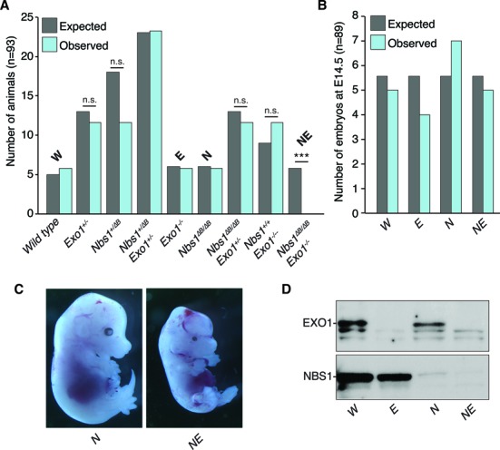
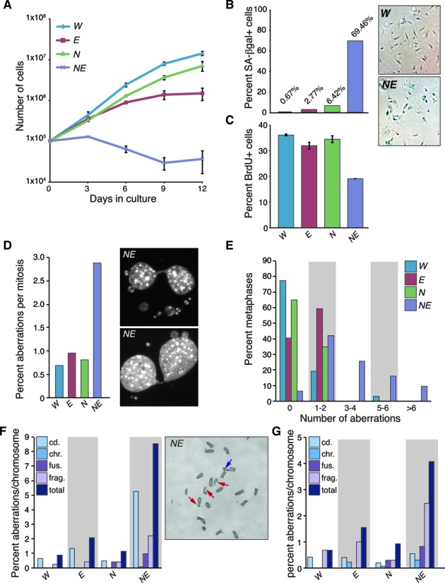
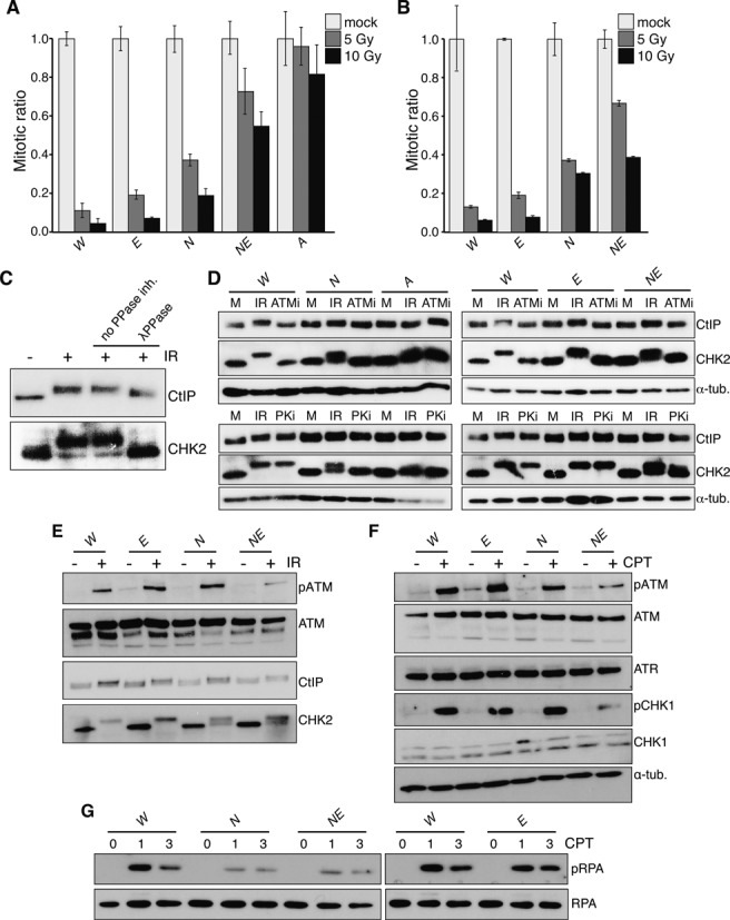
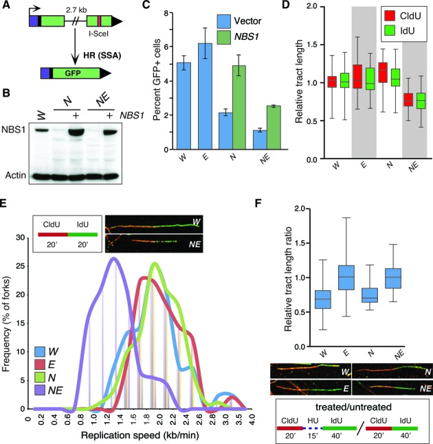
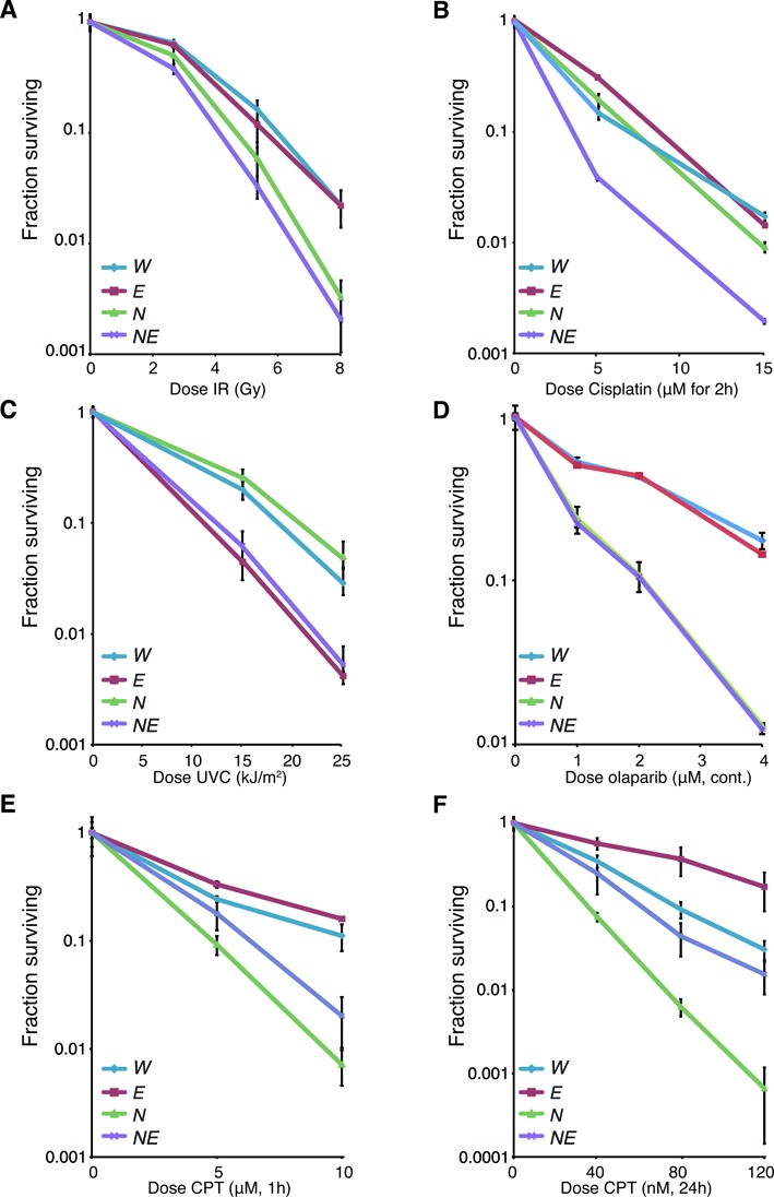
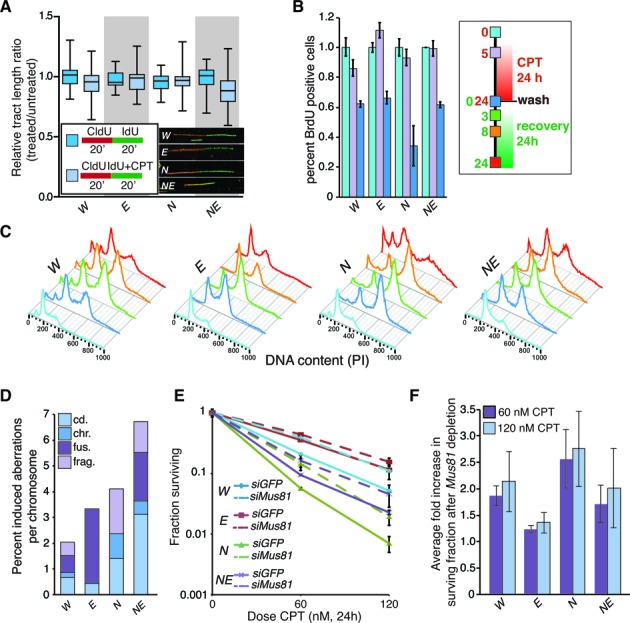
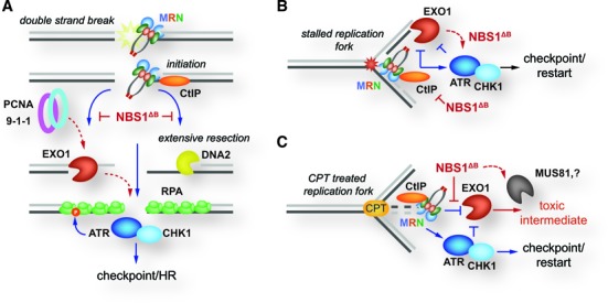
The maintenance of genome stability is critical for the suppression of diverse human pathologies that include developmental disorders, premature aging, infertility and predisposition to cancer. The DNA damage response (DDR) orchestrates the appropriate cellular responses following the detection of lesions to prevent genomic instability. The MRE11 complex is a sensor of DNA double strand breaks (DSBs) and plays key roles in multiple aspects of the DDR, including DNA end resection that is critical for signaling and DNA repair. The MRE11 complex has been shown to function both upstream and in concert with the 5'-3' exonuclease EXO1 in DNA resection, but it remains unclear to what extent EXO1 influences DSB responses independently of the MRE11 complex. Here we examine the genetic relationship of the MRE11 complex and EXO1 during mammalian development and in response to DNA damage. Deletion of Exo1 in mice expressing a hypomorphic allele of Nbs1 leads to severe developmental impairment, embryonic death and chromosomal instability. While EXO1 plays a minimal role in normal cells, its loss strongly influences DNA replication, DNA repair, checkpoint signaling and damage sensitivity in NBS1 hypomorphic cells. Collectively, our results establish a key role for EXO1 in modulating the severity of hypomorphic MRE11 complex mutations.


Figures

Figure 1.
Figure 1.
Deletion of Exo1 leads to embryonic lethality in hypomorphic Nbs1 mice. (A) Graph of expected and observed live born pups from double heterozygous breedings based on normal Mendelian inheritance (n = 93). For brevity, the primary genotypes are abbreviated as follows: wild type = W, Nbs1ΔB/ΔB = N, Exo1−/− = E, Nbs1ΔB/ΔBExo−/− = NE. Statistical analysis was performed using an unpaired t-test. ***P < 0.0001 and n.s. = not significant. (B) Graph of expected and observed E14.5 embryos from double heterozygous breedings based on normal Mendelian inheritance (n = 89). (C) Representative images of E14.5 embryos of the indicated genotype. (D) Western blotting of NBS1 and EXO1 from embryonic fibroblast cultures derived from E14.5 embryos.
Figure 2.
Figure 2.
EXO1 suppresses chromosomal instability in Nbs1 mutants. (A) Cell growth determined using a modified 3T3 protocol. Primary MEFs were counted and replated every 3 days and cumulative growth was plotted for the indicated genotypes. The genotypes are abbreviated throughout the figure as follows: wild type = W, Nbs1ΔB/ΔB = N, Exo1−/− = E, Nbs1ΔB/ΔBExo−/− = NE. (B) Premature senescence in NE double mutant cultures. Passage 6 MEFs were stained for senescence associated (SA) β-galactosidase activity and counted under magnification. The percentage of SA β-gal positive cells is plotted. (C) DNA synthesis is reduced in NE cell cultures. Passage 2 MEFs were pulsed with BrdU for 4 h and the percentage of BrdU positive cells was measured by flow cytometry combined with propidium iodide (PI) staining. (D) Increased anaphase bridges and micronuclei are evident in NE cultures. Quantification of the percentage of aberrant mitoses in primary cell cultures. A minimum of 100 mitoses are scored in each case. (E) Increased chromosomal instability in primary, early passage (p2-p3) NE cell cultures. The number of metaphase aberrations per metaphase were scored and binned as indicated in the figure. A higher percentage of NE metaphases show more than two aberrations per metaphase. (F) Plot of chromosomal aberration types (cd.=chromatid break, chr.=chromosome break, fus.=fusion, frag.=fragment) scored from multiple primary cell cultures depicted as percent aberrations per chromosome. NE cultures have particularly high numbers of chromatid breaks. Examples of aberrations scored in a partial NE metaphase are shown in the right panel. Red arrows indicate cd aberrations and blue arrow indicates a fusion. Additional examples are provided in Supplementary Figure S1. (G) Plot of chromosomal aberration types (as in (F)) identified in transformed cell cultures of the indicated genotypes. Total aberrations remain higher in NE cultures although chromosome fragments were the predominant lesion identified.
Figure 3.
Figure 3.
EXO1 is required for ATM/ATR signaling and the G2/M checkpoint in NBS1 mutant cells. (A) G2/M checkpoint activation in primary MEFs (p2-p3) 1 h after the indicated doses of IR scored by flow cytometric determination of the percentage of phospho-H3S10 positive cells with a G2 DNA content (determined by PI staining). The mitotic ratio is plotted (mock or IR treated cells, normalized to mock treated). The genotypes are abbreviated throughout the figure as follows: wild type = W, Nbs1ΔB/ΔB = N, Exo1−/− = E, Nbs1ΔB/ΔBExo−/− = NE and Atm−/− = A. (B) Checkpoint activation in transformed MEFs 1 hour after the indicated doses of IR, plotted as in (A). (C) The mobility shift of CHK2 and CtIP induced by IR is phosphorylation dependent. Lysates containing protease and phosphatase inhibitors were mock or IR treated and a mobility shift is evident for both proteins. Lysates from IR treated cells were harvested without phosphatase inhibitors (second 2 lanes) and treated with λ-phosphatase (last lane). (D) Hyperphosphorylation of CHK2 and CtIP is ATM dependent. Cell cultures of the indicated genotypes were pre-treated for 30 min with inhibitors of ATM (Atmi) or DNA-PKcs (PKi), mock or IR treated (10 Gy) in the presence of inhibitors and harvested 1 h post IR. The increased mobility of CHK2 and CtIP is lost in Atm−/− deficient cells or following the addition of ATMi. (E) Western blotting of the indicated proteins 1 h after 10 Gy IR treatment. (F) Western blotting of the indicated proteins 1 h after 1 mM CPT treatment. (G) Western blotting of the indicated proteins 1 and 3 h after 1 mM CPT treatment.
Figure 4.
Figure 4.
EXO1 influences DNA repair and DNA replication in Nbs1 mutants. (A) Schematic illustration of the SA-GFP based SSA assay. (B) Western blotting of cells transfected with or without a vector expressing human NBS1. The genotypes are abbreviated throughout the figure as follows: wild type = WNbs1ΔB/ΔB = NExo1−/− = E, and Nbs1ΔB/ΔB Exo−/− = NE. (C) Quantification of SSA mediated repair plotted as the percentage of GFP positive cells. Values for NE are corrected for the reduced percentage of cells in S/G2 determined by BrdU and PI staining (mean 58% compared to a mean of 73% in the other genotypes). The fold rescue with NBS1 expression is the same in N and NE (2.2 fold). (D) Measurement of replication tract lengths following CldU or IdU pulse labeling. (E) Calculation of replication fork velocity in the indicated genotypes (as described in ‘Materials and Methods’ section). Examples of representative forks from W and NE cultures are shown for comparison. (F) Assessment of replication fork restart following 1 mM HU treatment using the indicated scheme. Relative tract ratio is calculated by dividing the length of the IdU tract by that of the CldU tract ( = 2 under unperturbed conditions). Thus, higher values indicate faster restart following HU removal.
Figure 5.
Figure 5.
Differential effects of EXO1 loss on the damage sensitivity of Nbs1 mutants. Sensitivity of the cell cultures of the indicated genotypes to DNA damaging agents or PARP inhibitor (Olaparib) using the clonogenic survival assay. The genotypes are abbreviated in each case as follows: wild type = W, Nbs1ΔB/ΔB = N, Exo1−/− = E, and Nbs1ΔB/ΔB Exo−/− = NE. (A) IR sensitivity following treatment with 2, 5 or 8 Gy. (B) Cisplatin sensitivity following treatment at the indicated doses for 2 h. (C) UVC sensitivity after treatment with 15 or 25 J/m2. (D) Olaparib sensitivity following continuous treatment with the indicated doses. (E) CPT sensitivity (high) following treatment with the indicated doses for 1 h. (F) CPT sensitivity (low) following treatment with the indicated doses for 24 hours.
Figure 6.
Figure 6.
EXO1 modifies the cellular response to low dose camptothecin. (A) Measurement of replication fork tracts upon CPT treatment in the indicated genotypes. The tract length ratios (IdU/CldU) were not significantly different with or without 80 nM CPT treatment in the initial 20 min. The genotypes are abbreviated in each case as follows: wild type = WNbs1ΔB/ΔB = NExo1−/− = E, and Nbs1ΔB/ΔB Exo−/− = NE. (B) Analysis of BrdU incorporation at the indicated times post CPT addition by flow cytometry. The percentage of cells incorporating BrdU drops more dramatically in Nbs1ΔB/ΔB cultures after 24 hours of treatment compared to other genotypes. Time points (0, 5 and 24 h post CPT treatment) were assessed (legend corresponds to panels B and C). (C) Cell cycle profiles of cell cultures of the indicated genotypes following CPT treatment and withdrawal. (D) Metaphase aberrations induced by 50 nM CPT treatment and 4 h of recovery in the indicated genotypes. (E) Sensitivity of cell cultures of the indicated genotypes transfected with siRNA (siGFP as a control or 2 siRNAs against Mus81) to the indicated dose of camptothecin using the colony formation assay. Average results from triplicate experiments with 2 different Mus81 siRNAs (n = 6) are plotted and standard deviation indicated. (F) Average fold increase in survival following the depletion of MUS81 calculated by dividing the fraction surviving values of the MUS81 siRNA treated cells by the siGFP controls for each of the genotypes.
Figure 7.
Figure 7.
Proposed model of MRE11 complex and EXO1 interactions in replication and repair. (A) DSBs are recognized by the MRE11 complex (MRN) that initiates bidirectional resection through its nuclease activity. MRN promotes the exonuclease activity of both EXO1 and DNA2 that generate ssDNA that is bound by RPA. These RPA bound strands then promote the activation of ATR and CHK1 to promote checkpoint responses and homologous recombination. In cells expressing the NBS1ΔB protein, the ability of MRN to promote EXO1 and DNA2 mediated resection is reduced and RPA phosphorylation is impaired. Resection becomes dependent on EXO1 and it may be recruited via PCNA or the 9-1-1 complex to promote ATR/CHK1 activation and suppress more severe checkpoint signaling and repair defects. (B) Stalled replication forks are recognized by both MRN and EXO1 and excessive resection is suppressed by feedback from ATR and CHK1. In cells expressing the NBS1ΔB protein, EXO1 activity is increased, due to reduced negative feedback from ATR/CHK1, and it is able to compensate for defective MRN, thus suppressing more severe replication defects. (C) On forks that have stalled or regressed due to CPT, the combined actions of MRN and ATR/CHK1 may suppress the generation of intermediates by EXO1 that could be rendered cytotoxic by the actions of additional nucleases, such as MUS81.

References

    1. Jackson S.P., Bartek J. The DNA-damage response in human biology and disease. Nature. 2009;461:1071–1078. - PMC - PubMed
    1. Nussenzweig A., Nussenzweig M.C. Origin of chromosomal translocations in lymphoid cancer. Cell. 2010;141:27–38. - PMC - PubMed
    1. Halazonetis T.D., Gorgoulis V.G., Bartek J. An oncogene-induced DNA damage model for cancer development. Science. 2008;319:1352–1355. - PubMed
    1. Stracker T.H., Petrini J.H. The MRE11 complex: starting from the ends. Nat. Rev. Mol. Cell Biol. 2011;12:90–103. - PMC - PubMed
    1. Stracker T.H., Roig I., Knobel P.A., Marjanovic M. The ATM signaling network in development and disease. Front. Genet. 2013;4:37. - PMC - PubMed

Publication types

MeSH terms