. 2015 Aug 1;142(15):2633-40.
doi: 10.1242/dev.121707. Epub 2015 Jul 9.

Regulator of G-protein signaling 2 (RGS2) suppresses premature calcium release in mouse eggs

Affiliations

Regulator of G-protein signaling 2 (RGS2) suppresses premature calcium release in mouse eggs

Miranda L Bernhardt et al. Development. .

Abstract

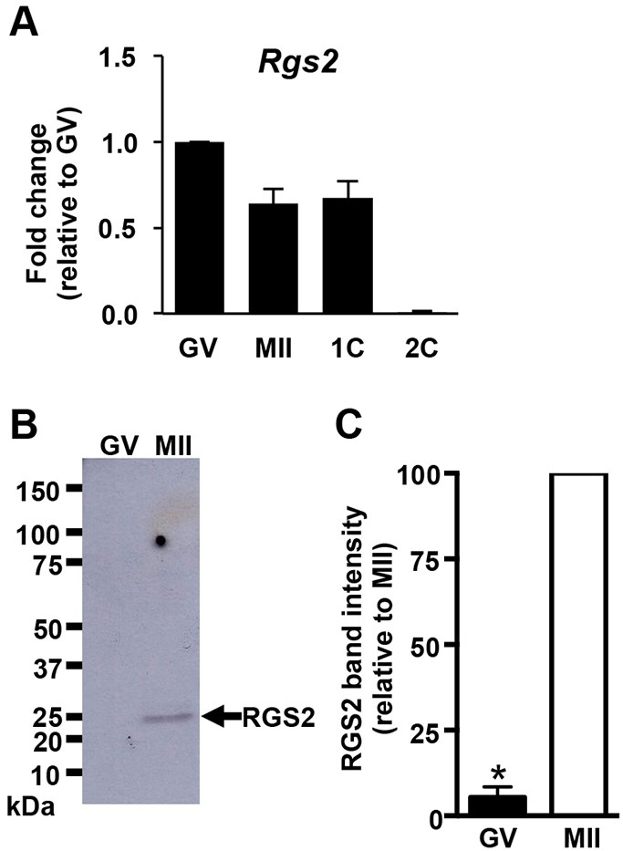
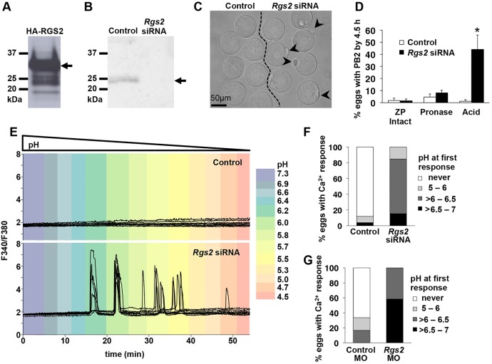
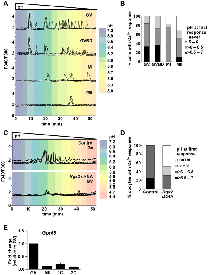
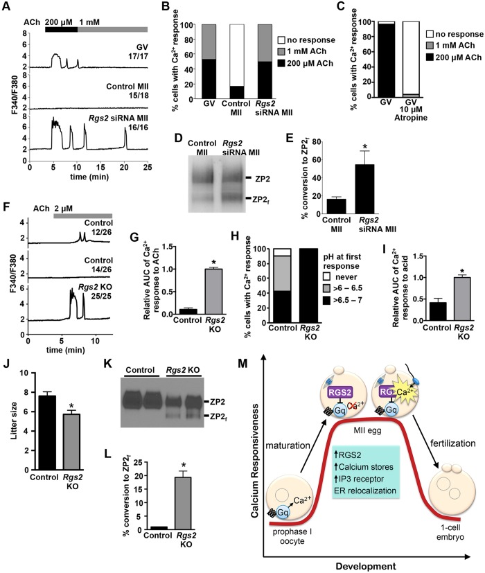
During oocyte maturation, capacity and sensitivity of Ca(2+) signaling machinery increases dramatically, preparing the metaphase II (MII)-arrested egg for fertilization. Upon sperm-egg fusion, Ca(2+) release from IP3-sensitive endoplasmic reticulum stores results in cytoplasmic Ca(2+) oscillations that drive egg activation and initiate early embryo development. Premature Ca(2+) release can cause parthenogenetic activation prior to fertilization; thus, preventing inappropriate Ca(2+) signaling is crucial for ensuring robust MII arrest. Here, we show that regulator of G-protein signaling 2 (RGS2) suppresses Ca(2+) release in MII eggs. Rgs2 mRNA was recruited for translation during oocyte maturation, resulting in ∼ 20-fold more RGS2 protein in MII eggs than in fully grown immature oocytes. Rgs2-siRNA-injected oocytes matured to MII; however, they had increased sensitivity to low pH and acetylcholine (ACh), which caused inappropriate Ca(2+) release and premature egg activation. When matured in vitro, RGS2-depleted eggs underwent spontaneous Ca(2+) increases that were sufficient to cause premature zona pellucida conversion. Rgs2(-/-) females had reduced litter sizes, and their eggs had increased sensitivity to low pH and ACh. Rgs2(-/-) eggs also underwent premature zona pellucida conversion in vivo. These findings indicate that RGS2 functions as a brake to suppress premature Ca(2+) release in eggs that are poised on the brink of development.

Keywords: Calcium; Egg activation; Gq; Meiotic maturation; Oocyte; RGS2.


Figures

Fig. 1.
Fig. 1.
RGS2 expression in oocytes, eggs and early embryos. (A) Rgs2 mRNA levels; all stages expressed relative to GV oocytes. N=3; graph shows mean±s.e.m. (B) Immunoblot of RGS2 protein in oocytes and eggs. Blot representative of 4 independent replicates; 50 oocytes or eggs/lane. (C) Quantitation of RGS2 immunoblot signal. N=4; graph shows mean±s.e.m. *P<0.05, Mann–Whitney test. GV, GV-stage oocytes; MII, MII eggs; 1C, 1-cell embryos; 2C, 2-cell embryos.
Fig. 2.
Fig. 2.
Acid-induced Ca2+ release causes resumption of meiosis in eggs lacking RGS2 protein. (A) Immunoblot of GV oocytes following microinjection with cRNA encoding HA-tagged RGS2, probed with monoclonal anti-RGS2 antibody. Arrow, full-length HA-RGS2 band. 20 oocytes/lane. (B) Immunoblot of RGS2 in control MII eggs or eggs matured to MII following microinjection at the GV stage with Rgs2 siRNA. 20 eggs/lane. Arrow, RGS2 band. (C) Appearance of siRNA-injected eggs following ZP removal with acid Tyrode's solution. Eggs in different groups are separated by dashed line. Arrowheads indicate second polar bodies. (D) Average percentage of eggs with second polar body (PB2) emitted by 4.5 h after the indicated treatment. N=5 independent replicates with 8-27 cells per group/replicate; graph shows mean±s.e.m. *P<0.05, ANOVA with Bonferroni's multiple comparison test. (E) Relative level of intracellular Ca2+ in response to lowering pH in control eggs or eggs lacking RGS2. Color indicates approximate pH at each time point. Eight representative tracings shown/group. (F) Percentage of siRNA-injected eggs with a rise in intracellular Ca2+ beginning at the indicated pH. (G) Percentage of morpholino (MO)-injected eggs with a rise in intracellular Ca2+ beginning at the indicated pH. Control MO, scrambled MO; Rgs2 MO, Rgs2-targeted MO. Graphs in F and G indicate cumulative percentage of 3-8 cells/group from n=4 or n=2 experiments, respectively.
Fig. 3.
Fig. 3.
RGS2 mediates loss of acid-induced Ca2+ response during oocyte maturation. (A) Relative level of intracellular Ca2+ in response to lowering pH in maturing oocytes. Graphs show 4-5 representative tracings/group. (B) Percentage of oocytes with a rise in intracellular Ca2+ beginning at the indicated pH. Graph indicates cumulative percentage of 2-5 cells/group from n=5 independent replicates. (C) Relative level of intracellular Ca2+ in response to lowering pH in control GV oocytes or GV oocytes overexpressing RGS2 (Rgs2 cRNA). Six representative tracings/group. (D) Percentage of oocytes with a rise in intracellular Ca2+ beginning at the indicated pH levels. Graph indicates cumulative percentage of 3-8 cells/group from n=4 independent replicates. (E) Gpr68 mRNA level; all stages expressed relative to GV oocytes. N=3; graph shows mean±s.e.m. GV, GV oocyte; GVBD, oocytes immediately following GV breakdown; MI, metaphase I stage; MII, MII eggs; 1C, 1-cell embryos; 2C, 2-cell embryos.
Fig. 4.
Fig. 4.
RGS2 inhibits acetylcholine (ACh)-induced Ca2+ release and premature ZP2 cleavage. (A) Relative level of intracellular Ca2+ in response to the indicated ACh concentrations. One representative tracing is shown per group, along with the proportion of cells displaying a similar pattern. GV, GV oocytes; Control MII, in vitro-matured MII eggs; Rgs2 siRNA MII, MII eggs matured in vitro following microinjection at the GV stage with Rgs2 siRNA. (B) Percentage of cells with a rise in intracellular Ca2+ beginning at the indicated ACh concentrations. Graph indicates cumulative percentage of 4-6 cells/group from n=4 independent replicates. (C) Effect of atropine on ACh-induced Ca2+ response in GV oocytes. Graph indicates cumulative percentage of 25 cells/group from n=3 independent replicates. (D) Immunoblot of ZP2 protein. Oocytes were microinjected with scrambled siRNA (control) or Rgs2 siRNA, then matured in vitro to MII. Blot represents 3 independent replicates; 12 eggs per lane. ZP2, full-length ZP2 protein; ZP2f, cleaved form of ZP2. (E) Quantitation of ZP2-to-ZP2f conversion. Graph shows mean±s.e.m. of 3 independent replicates. *P<0.05; t-test. (F) Relative level of intracellular Ca2+ in response to 2 µM ACh. Representative tracings are shown along with the proportion of cells displaying a similar pattern. Control, Rgs2+/+ eggs; Rgs2 KO, Rgs2−/− eggs. (G) Relative area under the curve (AUC) of ACh response. N=25-26 total eggs in 5 independent experiments. Graph shows mean±s.e.m. *P<0.05, t-test. (H) Number of eggs with a rise in intracellular Ca2+ beginning at the indicated pH. (I) Relative AUC of acid response. N=21-23 total eggs in 4 independent experiments. Graph shows mean±s.e.m. *P<0.05, t-test. (J) Average litter size for the indicated genotype. N=31-33 litters; *P<0.05, t-test. (K) Immunoblot of ZP2 protein from Rgs2+/+ (control) and Rgs2−/− (Rgs2 KO) eggs. Blot shows 2 of 3 replicates; 10 eggs per lane. (L) Quantitation of ZP2-to-ZP2f conversion in the indicated groups. Graph shows mean±s.e.m. of 3 replicates. *P<0.05; t-test. (M) Schematic summarizing RGS2 function after oocyte maturation in suppressing Ca2+ signaling mediated by Gq prior to fertilization.

Similar articles

Cited by

References

    1. Bernhardt M. L., Kim A. M., O'Halloran T. V. and Woodruff T. K. (2011). Zinc requirement during meiosis I-meiosis II transition in mouse oocytes is independent of the MOS-MAPK pathway. Biol. Reprod. 84, 526-536. 10.1095/biolreprod.110.086488 - DOI - PMC - PubMed
    1. Bernhardt M. L., Kong B. Y., Kim A. M., O'Halloran T. V. and Woodruff T. K. (2012). A zinc-dependent mechanism regulates meiotic progression in mammalian oocytes. Biol. Reprod. 86, 114 10.1095/biolreprod.111.097253 - DOI - PMC - PubMed
    1. Blake J. A., Bult C. J., Eppig J. T., Kadin J. A. and Richardson J. E. (2014). The Mouse Genome Database: integration of and access to knowledge about the laboratory mouse. Nucleic Acids Res. 42, D810-D817. 10.1093/nar/gkt1225 - DOI - PMC - PubMed
    1. Burkart A. D., Xiong B., Baibakov B., Jimenez-Movilla M. and Dean J. (2012). Ovastacin, a cortical granule protease, cleaves ZP2 in the zona pellucida to prevent polyspermy. J. Cell Biol. 197, 37-44. 10.1083/jcb.201112094 - DOI - PMC - PubMed
    1. Caratsch C., Eusebi F. and Salustri A. (1984). Acetylcholine receptors in monkey and rabbit oocytes. J. Cell. Physiol. 121, 415-418. 10.1002/jcp.1041210220 - DOI - PubMed

Publication types

LinkOut - more resources